 Movie
Movie Controller


+ Open data

- Basic information
| Entry | Database: PDB / ID: 4uem | ||||||
|---|---|---|---|---|---|---|---|
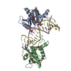
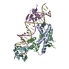
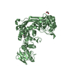
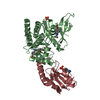
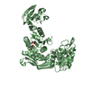
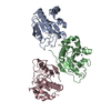
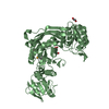
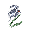
| Title | UCH-L5 in complex with the RPN13 DEUBAD domain | ||||||
 Components Components |
| ||||||
 Keywords Keywords |  HYDROLASE / DEUBIQUITINATING ENZYME / DUB / PROTEASOME HYDROLASE / DEUBIQUITINATING ENZYME / DUB / PROTEASOME | ||||||
| Function / homology |  Function and homology information Function and homology informationlateral ventricle development / regulation of DNA strand elongation / positive regulation of telomere maintenance in response to DNA damage / forebrain morphogenesis / Ino80 complex / cytosolic proteasome complex / proteasome regulatory particle, lid subcomplex / positive regulation of smoothened signaling pathway / midbrain development / regulation of chromosome organization ...lateral ventricle development / regulation of DNA strand elongation / positive regulation of telomere maintenance in response to DNA damage / forebrain morphogenesis / Ino80 complex / cytosolic proteasome complex / proteasome regulatory particle, lid subcomplex / positive regulation of smoothened signaling pathway / midbrain development / regulation of chromosome organization / endopeptidase inhibitor activity / molecular function inhibitor activity / proteasome binding / protein deubiquitination / regulation of DNA replication / regulation of embryonic development / endopeptidase activator activity / negative regulation of proteasomal ubiquitin-dependent protein catabolic process / proteasome assembly / regulation of DNA repair / regulation of proteasomal protein catabolic process / positive regulation of DNA repair / telomere maintenance / proteasome complex / Downregulation of TGF-beta receptor signaling / transcription elongation by RNA polymerase II / UCH proteinases / ubiquitin-dependent protein catabolic process / proteasome-mediated ubiquitin-dependent protein catabolic process / DNA recombination / protease binding / ubiquitinyl hydrolase 1 / cysteine-type deubiquitinase activity / Ub-specific processing proteases / regulation of cell cycle / chromatin remodeling / DNA repair / nucleolus / positive regulation of DNA-templated transcription / RNA binding / nucleoplasm / nucleus / plasma membrane / cytosol / cytoplasmSimilarity search - Function | ||||||
| Biological species |  HOMO SAPIENS (human) HOMO SAPIENS (human) | ||||||
| Method | X-RAY DIFFRACTION / SYNCHROTRON / MOLECULAR REPLACEMENT / Resolution: 2.82 Å | ||||||
 Authors Authors | Sahtoe, D.D. / Van Dijk, W.J. / El Oualid, F. / Ekkebus, R. / Ovaa, H. / Sixma, T.K. | ||||||
 Citation Citation | Journal: Mol.Cell / Year: 2015 Title: Mechanism of Uch-L5 Activation and Inhibition by Deubad Domains in Rpn13 and Ino80G. Authors: Sahtoe, D.D. / Van Dijk, W.J. / El Oualid, F. / Ekkebus, R. / Ovaa, H. / Sixma, T.K. | ||||||
| History |
|
- Structure visualization
| Structure viewer | Molecule: MolmilJmol/JSmol |
|---|
- Downloads & links
-Download
| PDBx/mmCIF format | 4uem.cif.gz | 167 KB | Display | PDBx/mmCIF format |
|---|---|---|---|---|
| PDB format | pdb4uem.ent.gz | 133.6 KB | Display | PDB format |
| PDBx/mmJSON format | 4uem.json.gz | Tree view | PDBx/mmJSON format | |
| Others |  Other downloads Other downloads |
-Validation report
| Arichive directory | https://data.pdbj.org/pub/pdb/validation_reports/ue/4uemftp://data.pdbj.org/pub/pdb/validation_reports/ue/4uem | HTTPS FTP |
|---|
-Related structure data
| Related structure data |  4uelC 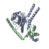 4uf5C 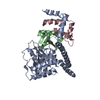 4uf6C 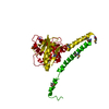 3ihrS C: citing same article ( S: Starting model for refinement |
|---|---|
| Similar structure data |


-Links
 PDBj
PDBj


- Assembly
| Deposited unit | 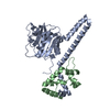
| ||||||||
|---|---|---|---|---|---|---|---|---|---|
| 1 |
| ||||||||
| Unit cell |
|
-Components
| #1: Protein | Mass: 37734.934 Da / Num. of mol.: 1 Source method: isolated from a genetically manipulated source Details: ISOFORM 3 / Source: (gene. exp.) HOMO SAPIENS (human) / Plasmid: PGEX-NKI-3C-LIC / Production host:  ESCHERICHIA COLI (E. coli) / Strain (production host): BL21(DE3) / Variant (production host): ROSETTA 2 / References: UniProt: Q9Y5K5, ubiquitinyl hydrolase 1 ESCHERICHIA COLI (E. coli) / Strain (production host): BL21(DE3) / Variant (production host): ROSETTA 2 / References: UniProt: Q9Y5K5, ubiquitinyl hydrolase 1 |
|---|---|
| #2: Protein | Mass: 13065.674 Da / Num. of mol.: 1 / Fragment: DEUBAD DOMAIN, RESIDUES 266-388 Source method: isolated from a genetically manipulated source Source: (gene. exp.) HOMO SAPIENS (human) / Plasmid: PCDF-NKI-HIS-3C-LIC / Production host: ESCHERICHIA COLI (E. coli) / Strain (production host): BL21(DE3) / Variant (production host): ROSETTA 2 / References: UniProt: Q16186 |
| Sequence details | ISOFORM 3 |
-Experimental details
-Experiment
| Experiment | Method: X-RAY DIFFRACTION |
|---|
- Sample preparation
| Crystal | Density Matthews: 2.7 Å3/Da / Density % sol: 54 % / Description: NONE |
|---|---|
| Crystal grow | Temperature: 277 K Details: 100 MM BIS-TRIS-PROPANE PH 6.4, 230 MM NABR, 21% PEG3350. 4 DEGREES CELSIUS |
-Data collection
| Diffraction | Mean temperature: 100 K |
|---|---|
| Diffraction source | Source: SYNCHROTRON / Site: ESRF  / Beamline: ID14-4 / Wavelength: 0.9793 / Beamline: ID14-4 / Wavelength: 0.9793 |
| Detector | Type: ADSC CCD / Detector: CCD / Date: Jul 25, 2013 |
| Radiation | Protocol: SINGLE WAVELENGTH / Monochromatic (M) / Laue (L): M / Scattering type: x-ray |
| Radiation wavelength | Wavelength: 0.9793 Å / Relative weight: 1 |
| Reflection | Resolution: 2.82→33.54 Å / Num. obs: 13635 / % possible obs: 98.5 % / Observed criterion σ(I): 2.2 / Redundancy: 3 % / Biso Wilson estimate: 77.01 Å2 / Rmerge(I) obs: 0.06 / Net I/σ(I): 15.3 |
| Reflection shell | Resolution: 2.82→2.97 Å / Redundancy: 3.7 % / Rmerge(I) obs: 0.86 / Mean I/σ(I) obs: 2.2 / % possible all: 97.3 |
- Processing
| Software |
| ||||||||||||||||||||||||||||||||||||||||||||||||||||||||||||||||||||||||||||||||||||||||||||||||||||||||||||||||||||||||||||||||||||||||||||||||||||||
|---|---|---|---|---|---|---|---|---|---|---|---|---|---|---|---|---|---|---|---|---|---|---|---|---|---|---|---|---|---|---|---|---|---|---|---|---|---|---|---|---|---|---|---|---|---|---|---|---|---|---|---|---|---|---|---|---|---|---|---|---|---|---|---|---|---|---|---|---|---|---|---|---|---|---|---|---|---|---|---|---|---|---|---|---|---|---|---|---|---|---|---|---|---|---|---|---|---|---|---|---|---|---|---|---|---|---|---|---|---|---|---|---|---|---|---|---|---|---|---|---|---|---|---|---|---|---|---|---|---|---|---|---|---|---|---|---|---|---|---|---|---|---|---|---|---|---|---|---|---|---|---|
| Refinement | Method to determine structure: MOLECULAR REPLACEMENT Starting model: PDB ENTRY 3IHR Resolution: 2.82→28.853 Å / SU ML: 0.48 / σ(F): 1.34 / Phase error: 31.33 / Stereochemistry target values: ML
| ||||||||||||||||||||||||||||||||||||||||||||||||||||||||||||||||||||||||||||||||||||||||||||||||||||||||||||||||||||||||||||||||||||||||||||||||||||||
| Solvent computation | Shrinkage radii: 0.9 Å / VDW probe radii: 1.11 Å / Solvent model: FLAT BULK SOLVENT MODEL | ||||||||||||||||||||||||||||||||||||||||||||||||||||||||||||||||||||||||||||||||||||||||||||||||||||||||||||||||||||||||||||||||||||||||||||||||||||||
| Refinement step | Cycle: LAST / Resolution: 2.82→28.853 Å
| ||||||||||||||||||||||||||||||||||||||||||||||||||||||||||||||||||||||||||||||||||||||||||||||||||||||||||||||||||||||||||||||||||||||||||||||||||||||
| Refine LS restraints |
| ||||||||||||||||||||||||||||||||||||||||||||||||||||||||||||||||||||||||||||||||||||||||||||||||||||||||||||||||||||||||||||||||||||||||||||||||||||||
| LS refinement shell |
| ||||||||||||||||||||||||||||||||||||||||||||||||||||||||||||||||||||||||||||||||||||||||||||||||||||||||||||||||||||||||||||||||||||||||||||||||||||||
| Refinement TLS params. | Method: refined / Refine-ID: X-RAY DIFFRACTION
| ||||||||||||||||||||||||||||||||||||||||||||||||||||||||||||||||||||||||||||||||||||||||||||||||||||||||||||||||||||||||||||||||||||||||||||||||||||||
| Refinement TLS group |
|
