 Movie
Movie Controller


+ Open data

- Basic information
| Entry | Database: PDB / ID: 4ued | ||||||
|---|---|---|---|---|---|---|---|
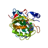
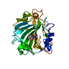
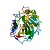
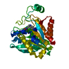
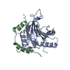
| Title | Complex of human eIF4E with the 4E binding protein 4E-BP1 | ||||||
 Components Components |
| ||||||
 Keywords Keywords |  TRANSLATION / GENE REGULATION / CAP BINDING PROTEIN / 4E BINDING PROTEIN / TRANSLATIONAL REPRESSION TRANSLATION / GENE REGULATION / CAP BINDING PROTEIN / 4E BINDING PROTEIN / TRANSLATIONAL REPRESSION | ||||||
| Function / homology |  Function and homology information Function and homology informationActivation of the mRNA upon binding of the cap-binding complex and eIFs, and subsequent binding to 43S / : / eukaryotic initiation factor 4G binding / eukaryotic initiation factor 4E binding / regulation of translation at postsynapse, modulating synaptic transmission / RNA cap binding / chromatoid body / eukaryotic translation initiation factor 4F complex / Z-decay: degradation of maternal mRNAs by zygotically expressed factors / mRNA cap binding ...Activation of the mRNA upon binding of the cap-binding complex and eIFs, and subsequent binding to 43S / : / eukaryotic initiation factor 4G binding / eukaryotic initiation factor 4E binding / regulation of translation at postsynapse, modulating synaptic transmission / RNA cap binding / chromatoid body / eukaryotic translation initiation factor 4F complex / Z-decay: degradation of maternal mRNAs by zygotically expressed factors / mRNA cap binding / Deadenylation of mRNA / Transport of the SLBP independent Mature mRNA / RNA 7-methylguanosine cap binding / Transport of the SLBP Dependant Mature mRNA / M-decay: degradation of maternal mRNAs by maternally stored factors / Transport of Mature mRNA Derived from an Intronless Transcript / RISC complex / Ribosomal scanning and start codon recognition / Translation initiation complex formation / stem cell population maintenance / mTORC1-mediated signalling / GTP hydrolysis and joining of the 60S ribosomal subunit / negative regulation of neuron differentiation / TOR signaling / L13a-mediated translational silencing of Ceruloplasmin expression / behavioral fear response / mRNA export from nucleus / translation repressor activity / negative regulation of translational initiation / translational initiation / translation initiation factor binding / translation initiation factor activity / positive regulation of mitotic cell cycle / cellular response to dexamethasone stimulus / P-body / neuron differentiation / G1/S transition of mitotic cell cycle / ISG15 antiviral mechanism / cytoplasmic stress granule / cytoplasmic ribonucleoprotein granule / regulation of translation / postsynapse / DNA-binding transcription factor binding / negative regulation of translation / nuclear speck / glutamatergic synapse / perinuclear region of cytoplasm / enzyme binding / RNA binding / extracellular exosome / nucleus / cytosol / cytoplasmSimilarity search - Function | ||||||
| Biological species |  HOMO SAPIENS (human) HOMO SAPIENS (human) | ||||||
| Method | X-RAY DIFFRACTION / SYNCHROTRON / MOLECULAR REPLACEMENT / Resolution: 1.75 Å | ||||||
 Authors Authors | Peter, D. / Weichenrieder, O. | ||||||
 Citation Citation | Journal: Mol.Cell / Year: 2015 Title: Molecular Architecture of 4E-BP Translational Inhibitors Bound to Eif4E. Authors: Peter, D. / Igreja, C. / Weber, R. / Wohlbold, L. / Weiler, C. / Ebertsch, L. / Weichenrieder, O. / Izaurralde, E. | ||||||
| History |
| ||||||
| Remark 650 | HELIX DETERMINATION METHOD: AUTHOR PROVIDED. |
- Structure visualization
| Structure viewer | Molecule: MolmilJmol/JSmol |
|---|
- Downloads & links
-Download
| PDBx/mmCIF format | 4ued.cif.gz | 138.5 KB | Display | PDBx/mmCIF format |
|---|---|---|---|---|
| PDB format | pdb4ued.ent.gz | 111.3 KB | Display | PDB format |
| PDBx/mmJSON format | 4ued.json.gz | Tree view | PDBx/mmJSON format | |
| Others |  Other downloads Other downloads |
-Validation report
| Arichive directory | https://data.pdbj.org/pub/pdb/validation_reports/ue/4uedftp://data.pdbj.org/pub/pdb/validation_reports/ue/4ued | HTTPS FTP |
|---|
-Related structure data
| Related structure data | 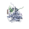 4ue8C 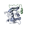 4ue9C 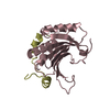 4ueaC 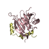 4uebC 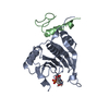 4uecC 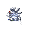 4tpwS C: citing same article ( S: Starting model for refinement |
|---|---|
| Similar structure data |


-Links
 PDBj
PDBj


- Assembly
| Deposited unit | 
| ||||||||
|---|---|---|---|---|---|---|---|---|---|
| 1 |
| ||||||||
| Unit cell |
|
-Components
| #1: Protein | EIF4E / EIF4E / EIF-4F 25 KDA SUBUNIT / MRNA CAP-BINDING PROTEIN Mass: 21657.625 Da / Num. of mol.: 1 / Fragment: RESIDUES 36-217 Source method: isolated from a genetically manipulated source Source: (gene. exp.) HOMO SAPIENS (human) / Plasmid: PETMCN (PNYC) / Production host:  ESCHERICHIA COLI (E. coli) / Strain (production host): BL21(DE3) / Variant (production host): STAR / References: UniProt: P06730 ESCHERICHIA COLI (E. coli) / Strain (production host): BL21(DE3) / Variant (production host): STAR / References: UniProt: P06730 |
|---|---|
| #2: Protein/peptide | Mass: 4333.087 Da / Num. of mol.: 1 / Fragment: RESIDUES 50-83 Source method: isolated from a genetically manipulated source Source: (gene. exp.) HOMO SAPIENS (human) / Plasmid: PETMCN (PNEA) / Production host: ESCHERICHIA COLI (E. coli) / Strain (production host): BL21(DE3) / Variant (production host): STAR / References: UniProt: Q13541 |
| #3: Water | ChemComp-HOH / Water Mass: 18.015 Da / Num. of mol.: 119 / Source method: isolated from a natural source / Formula: H2O Mass: 18.015 Da / Num. of mol.: 119 / Source method: isolated from a natural source / Formula: H2O |
| Sequence details | THE FIRST FOUR RESIDUES OF CHAIN A REMAIN FROM THE EXPRESSION TAG. THE FIRST FOUR RESIDUES OF CHAIN ...THE FIRST FOUR RESIDUES OF CHAIN A REMAIN FROM THE EXPRESSION |
-Experimental details
-Experiment
| Experiment | Method: X-RAY DIFFRACTION / Number of used crystals: 1 |
|---|
- Sample preparation
| Crystal | Density Matthews: 2.1 Å3/Da / Density % sol: 42 % / Description: NONE |
|---|---|
| Crystal grow | pH: 8.5 Details: 0.03M EACH DIETHYLENE GLYCOL, TRIETHYLENE GLYCOL, TETRAETHYLENE GLYCOL, PENTAETHYLENE GLYCOL, 0.1M BICINE/TRIZMA BASE PH 8.5, 12.5% PEG1000, 12.5% PEG3350, 12.5% MPD |
-Data collection
| Diffraction | Mean temperature: 100 K |
|---|---|
| Diffraction source | Source: SYNCHROTRON / Site: SLS  / Beamline: X10SA / Wavelength: 1.07 / Beamline: X10SA / Wavelength: 1.07 |
| Detector | Type: DECTRIS PILATUS 6M / Detector: PIXEL / Date: Aug 25, 2014 / Details: DYNAMICALLY BENDABLE MIRRORS |
| Radiation | Monochromator: SI(111) / Protocol: SINGLE WAVELENGTH / Monochromatic (M) / Laue (L): M / Scattering type: x-ray |
| Radiation wavelength | Wavelength: 1.07 Å / Relative weight: 1 |
| Reflection | Resolution: 1.75→37.9 Å / Num. obs: 20595 / % possible obs: 96.9 % / Observed criterion σ(I): -3 / Redundancy: 2.9 % / Biso Wilson estimate: 24.27 Å2 / Rsym value: 0.04 / Net I/σ(I): 13.4 |
| Reflection shell | Resolution: 1.75→1.8 Å / Redundancy: 2.8 % / Mean I/σ(I) obs: 2.5 / Rsym value: 0.31 / % possible all: 98.1 |
- Processing
| Software |
| |||||||||||||||||||||||||||||||||||||||||||||||||||||||||||||||
|---|---|---|---|---|---|---|---|---|---|---|---|---|---|---|---|---|---|---|---|---|---|---|---|---|---|---|---|---|---|---|---|---|---|---|---|---|---|---|---|---|---|---|---|---|---|---|---|---|---|---|---|---|---|---|---|---|---|---|---|---|---|---|---|---|
| Refinement | Method to determine structure: MOLECULAR REPLACEMENT Starting model: PDB ENTRY 4TPW CHAIN A Resolution: 1.75→37.906 Å / SU ML: 0.2 / σ(F): 1.36 / Phase error: 24.46 / Stereochemistry target values: ML Details: HYDROGENS WERE REFINED IN THE RIDING POSITIONS. THE SIDECHAINS OF THE FOLLOWING RESIDUES WERE TRUNCATED AT C-BETA ATOMS. CHAIN B, RESIDUES 73, 74. THE FOLLOWING RESIDUES WERE MODELED AS ...Details: HYDROGENS WERE REFINED IN THE RIDING POSITIONS. THE SIDECHAINS OF THE FOLLOWING RESIDUES WERE TRUNCATED AT C-BETA ATOMS. CHAIN B, RESIDUES 73, 74. THE FOLLOWING RESIDUES WERE MODELED AS DOUBLE CONFORMATIONS. CHAIN A, RESIDUES 64, 92, 172. THE FOLLOWING RESIDUES ARE DISORDERED. CHAIN A, RESIDUES 205 TO 211.
| |||||||||||||||||||||||||||||||||||||||||||||||||||||||||||||||
| Solvent computation | Shrinkage radii: 0.9 Å / VDW probe radii: 1.11 Å / Solvent model: FLAT BULK SOLVENT MODEL | |||||||||||||||||||||||||||||||||||||||||||||||||||||||||||||||
| Displacement parameters | Biso mean: 32.8 Å2 | |||||||||||||||||||||||||||||||||||||||||||||||||||||||||||||||
| Refinement step | Cycle: LAST / Resolution: 1.75→37.906 Å
| |||||||||||||||||||||||||||||||||||||||||||||||||||||||||||||||
| Refine LS restraints |
| |||||||||||||||||||||||||||||||||||||||||||||||||||||||||||||||
| LS refinement shell |
|
