 Movie
Movie Controller

[English] 日本語
 Yorodumi
- PDB-4ue4: Structural basis for targeting and elongation arrest of Bacillus ... -
+ Open data

- Basic information
| Entry | Database: PDB / ID: 4ue4 | ||||||
|---|---|---|---|---|---|---|---|
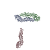
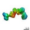
| Title | Structural basis for targeting and elongation arrest of Bacillus signal recognition particle | ||||||
 Components Components |
| ||||||
 Keywords Keywords |  TRANSLATION / SIGNAL RECOGNITION PARTICLE (SRP) / STALLED-RIBOSOME / TRANSLOCATION / MIFM STALLING TRANSLATION / SIGNAL RECOGNITION PARTICLE (SRP) / STALLED-RIBOSOME / TRANSLOCATION / MIFM STALLING | ||||||
| Function / homology |  Function and homology information Function and homology informationFtsQBL complex / divisome complex / signal recognition particle / signal-recognition-particle GTPase / 7S RNA binding / SRP-dependent cotranslational protein targeting to membrane / FtsZ-dependent cytokinesis / division septum assembly / cell division site / cell division ...FtsQBL complex / divisome complex / signal recognition particle / signal-recognition-particle GTPase / 7S RNA binding / SRP-dependent cotranslational protein targeting to membrane / FtsZ-dependent cytokinesis / division septum assembly / cell division site / cell division / GTPase activity / GTP binding / ATP hydrolysis activity / identical protein binding / plasma membraneSimilarity search - Function | ||||||
| Biological species |  BACILLUS SUBTILIS SUBSP. SUBTILIS STR. 168 (bacteria) BACILLUS SUBTILIS SUBSP. SUBTILIS STR. 168 (bacteria) | ||||||
| Method | ELECTRON MICROSCOPY / single particle reconstruction / cryo EM / Resolution: 7 Å | ||||||
 Authors Authors | Beckert, B. / Kedrov, A. / Sohmen, D. / Kempf, G. / Wild, K. / Sinning, I. / Stahlberg, H. / Wilson, D.N. / Beckmann, R. | ||||||
 Citation Citation | Journal: Nat Struct Mol Biol / Year: 2015 Title: Translational arrest by a prokaryotic signal recognition particle is mediated by RNA interactions. Authors: Bertrand Beckert / Alexej Kedrov / Daniel Sohmen / Georg Kempf / Klemens Wild / Irmgard Sinning / Henning Stahlberg / Daniel N Wilson / Roland Beckmann /   Abstract: The signal recognition particle (SRP) recognizes signal sequences of nascent polypeptides and targets ribosome-nascent chain complexes to membrane translocation sites. In eukaryotes, translating ...The signal recognition particle (SRP) recognizes signal sequences of nascent polypeptides and targets ribosome-nascent chain complexes to membrane translocation sites. In eukaryotes, translating ribosomes are slowed down by the Alu domain of SRP to allow efficient targeting. In prokaryotes, however, little is known about the structure and function of Alu domain-containing SRPs. Here, we report a complete molecular model of SRP from the Gram-positive bacterium Bacillus subtilis, based on cryo-EM. The SRP comprises two subunits, 6S RNA and SRP54 or Ffh, and it facilitates elongation slowdown similarly to its eukaryotic counterpart. However, protein contacts with the small ribosomal subunit observed for the mammalian Alu domain are substituted in bacteria by RNA-RNA interactions of 6S RNA with the α-sarcin-ricin loop and helices H43 and H44 of 23S rRNA. Our findings provide a structural basis for cotranslational targeting and RNA-driven elongation arrest in prokaryotes. | ||||||
| History |
|
- Structure visualization
| Movie |
Movie viewer |
|---|---|
| Structure viewer | Molecule: MolmilJmol/JSmol |
- Downloads & links
-Download
| PDBx/mmCIF format | 4ue4.cif.gz | 157.9 KB | Display | PDBx/mmCIF format |
|---|---|---|---|---|
| PDB format | pdb4ue4.ent.gz | 110.4 KB | Display | PDB format |
| PDBx/mmJSON format | 4ue4.json.gz | Tree view | PDBx/mmJSON format | |
| Others |  Other downloads Other downloads |
-Validation report
| Arichive directory | https://data.pdbj.org/pub/pdb/validation_reports/ue/4ue4ftp://data.pdbj.org/pub/pdb/validation_reports/ue/4ue4 | HTTPS FTP |
|---|
-Related structure data
| Related structure data | 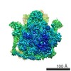 2843MC 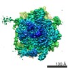 2844C 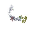 4ue5C C: citing same article ( M: map data used to model this data |
|---|---|
| Similar structure data |
-Links
 PDBj
PDBj


- Assembly
| Deposited unit | 
|
|---|---|
| 1 |
|
-Components
| #1: RNA chain | Mass: 85909.688 Da / Num. of mol.: 1 / Source method: isolated from a natural source Source: (natural) BACILLUS SUBTILIS SUBSP. SUBTILIS STR. 168 (bacteria)References: GenBank: 40130 |
|---|---|
| #2: Protein/peptide | Mass: 2466.997 Da / Num. of mol.: 1 / Source method: isolated from a natural source Source: (natural) BACILLUS SUBTILIS SUBSP. SUBTILIS STR. 168 (bacteria)References: UniProt: P06136*PLUS |
| #3: Protein | / FIFTY-FOUR HOMOLOG / FFH Mass: 11660.719 Da / Num. of mol.: 1 / Fragment: M DOMAIN Source method: isolated from a genetically manipulated source Source: (gene. exp.) BACILLUS SUBTILIS SUBSP. SUBTILIS STR. 168 (bacteria)Production host: ESCHERICHIA COLI (E. coli) / Strain (production host): BL21 / References: UniProt: P37105 |
-Experimental details
-Experiment
| Experiment | Method: ELECTRON MICROSCOPY |
|---|---|
| EM experiment | Aggregation state: PARTICLE / 3D reconstruction method: single particle reconstruction |
- Sample preparation
| Component | Name: BACILLUS SUBTILIS MIFM STALLED RIBOSOME IN COMPLEX WITH SIGNAL RECOGNITION PARTICLE (SRP) Type: RIBOSOME |
|---|---|
| Buffer solution | Name: HEPERS 20 MM, KOAC 150 MM, MG(OAC)2 10MM, NIKKOL 0.005%, DTT 1MM, 2% GLYCEROL pH: 7.6 Details: HEPERS 20 MM, KOAC 150 MM, MG(OAC)2 10MM, NIKKOL 0.005%, DTT 1MM, 2% GLYCEROL |
| Specimen | Embedding applied: NO / Shadowing applied: NO / Staining applied: NO / Vitrification applied: YES |
| Specimen support | Details: CARBON |
| Vitrification | Instrument: FEI VITROBOT MARK IV / Cryogen name: ETHANE Details: VITRIFICATION 1 -- CRYOGEN- ETHANE, INSTRUMENT- FEI VITROBOT MARK IV, |
- Electron microscopy imaging
| Experimental equipment |  Model: Titan Krios / Image courtesy: FEI Company |
|---|---|
| Microscopy | Model: FEI TITAN KRIOS / Date: Dec 10, 2013 |
| Electron gun | Electron source: FIELD EMISSION GUN / Accelerating voltage: 300 kV / Illumination mode: SPOT SCAN |
| Electron lens | Mode: BRIGHT FIELDBright-field microscopy / Nominal defocus max: 4000 nm / Nominal defocus min: 800 nm / Cs: 2.7 mm |
| Image recording | Electron dose: 20 e/Å2 / Film or detector model: TVIPS TEMCAM-F816 (8k x 8k) |
- Processing
| EM software | Name: SPIDER / Category: 3D reconstruction | ||||||||||||
|---|---|---|---|---|---|---|---|---|---|---|---|---|---|
| CTF correction | Details: MICROGRAPH | ||||||||||||
| Symmetry | Point symmetry: C1 (asymmetric) | ||||||||||||
| 3D reconstruction | Method: PROJECTION MATCHING / Resolution: 7 Å / Num. of particles: 75900 / Actual pixel size: 0.953 Å Details: SUBMISSION BASED ON EXPERIMENTAL DATA FROM EMDB EMD-2843. (DEPOSITION ID: 12993). Symmetry type: POINT | ||||||||||||
| Atomic model building | Protocol: RIGID BODY FIT / Details: METHOD--RIGID BODY | ||||||||||||
| Refinement | Highest resolution: 7 Å | ||||||||||||
| Refinement step | Cycle: LAST / Highest resolution: 7 Å
|
