 Movie
Movie Controller

[English] 日本語
 Yorodumi
- PDB-4udi: Crystal structure of b-1,4-mannopyranosyl-chitobiose phosphorylas... -
+ Open data

- Basic information
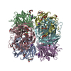
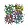
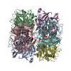
| Entry | Database: PDB / ID: 4udi | ||||||
|---|---|---|---|---|---|---|---|
| Title | Crystal structure of b-1,4-mannopyranosyl-chitobiose phosphorylase at 1.85 Angstrom from unknown human gut bacteria (Uhgb_MP) | ||||||
 Components Components | UHGB_MP | ||||||
 Keywords Keywords |  TRANSFERASE / GLYCOSIDE HYDROLASE FAMILY 130 / B-1 / 4-MANNOPYRANOSYL-CHITOBIOSE PHOSPHORYLASE / N-GLYCAN PHOSPHOROLYSIS TRANSFERASE / GLYCOSIDE HYDROLASE FAMILY 130 / B-1 / 4-MANNOPYRANOSYL-CHITOBIOSE PHOSPHORYLASE / N-GLYCAN PHOSPHOROLYSIS | ||||||
| Function / homology |  Function and homology information Function and homology information | ||||||
| Biological species | UNCULTURED ORGANISM (environmental samples) | ||||||
| Method | X-RAY DIFFRACTION / SYNCHROTRON / MOLECULAR REPLACEMENT / Resolution: 1.8 Å | ||||||
 Authors Authors | Ladeveze, S. / Cioci, G. / Potocki-Veronese, G. / Tranier, S. / Mourey, L. | ||||||
 Citation Citation | Journal: Acta Crystallogr.,Sect.D / Year: 2015 Title: Structural Bases for N-Glycan Processing by Mannoside Phosphorylase. Authors: Ladeveze, S. / Cioci, G. / Roblin, P. / Mourey, L. / Tranier, S. / Potocki-Veronese, G. #1: Journal: J.Biol.Chem. / Year: 2013 Title: Role of Glycoside Phosphorylases in Mannose Foraging by Human Gut Bacteria. Authors: Ladeveze, S. / Tarquis, L. / Cecchini, D.A. / Bercovici, J. / Andre, I. / Topham, C.M. / Morel, S. / Laville, E. / Monsan, P. / Lombard, V. / Henrissat, B. / Potocki-Veronese, G. | ||||||
| History |
|
- Structure visualization
| Structure viewer | Molecule: MolmilJmol/JSmol |
|---|
- Downloads & links
-Download
| PDBx/mmCIF format | 4udi.cif.gz | 822.6 KB | Display | PDBx/mmCIF format |
|---|---|---|---|---|
| PDB format | pdb4udi.ent.gz | 685.2 KB | Display | PDB format |
| PDBx/mmJSON format | 4udi.json.gz | Tree view | PDBx/mmJSON format | |
| Others |  Other downloads Other downloads |
-Validation report
| Arichive directory | https://data.pdbj.org/pub/pdb/validation_reports/ud/4udiftp://data.pdbj.org/pub/pdb/validation_reports/ud/4udi | HTTPS FTP |
|---|
-Related structure data
| Related structure data | 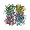 4udgC  4udjC  4udkC  1vkdS C: citing same article ( S: Starting model for refinement |
|---|---|
| Similar structure data |


-Links
 PDBj
PDBj- Assembly
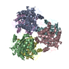
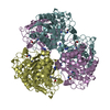
| Deposited unit | 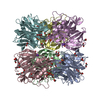
| ||||||||
|---|---|---|---|---|---|---|---|---|---|
| 1 |
| ||||||||
| Unit cell |
|
-Components
-Protein , 1 types, 6 molecules ABCDEF
| #1: Protein | Mass: 39321.422 Da / Num. of mol.: 6 Source method: isolated from a genetically manipulated source Source: (gene. exp.) UNCULTURED ORGANISM (environmental samples) Description: FECAL SAMPLE FROM HOMO SAPIENS / Plasmid: PET28A / Production host:  ESCHERICHIA COLI (E. coli) / Strain (production host): BL21 / Variant (production host): AI ESCHERICHIA COLI (E. coli) / Strain (production host): BL21 / Variant (production host): AIReferences: UniProt: D9ZDQ9, Transferases; Glycosyltransferases; Hexosyltransferases |
|---|
-Non-polymers , 6 types, 1126 molecules 


| #2: Chemical | ChemComp-PO4 / Phosphate Mass: 94.971 Da / Num. of mol.: 6 / Source method: obtained synthetically / Formula: PO4 Mass: 94.971 Da / Num. of mol.: 6 / Source method: obtained synthetically / Formula: PO4#3: Chemical | ChemComp-GOL / Glycerol Mass: 92.094 Da / Num. of mol.: 27 / Source method: obtained synthetically / Formula: C3H8O3 Mass: 92.094 Da / Num. of mol.: 27 / Source method: obtained synthetically / Formula: C3H8O3#4: Chemical | ChemComp-K /  Mass: 39.098 Da / Num. of mol.: 6 / Source method: obtained synthetically / Formula: K Mass: 39.098 Da / Num. of mol.: 6 / Source method: obtained synthetically / Formula: K#5: Chemical | ChemComp-EDO / Ethylene glycol Mass: 62.068 Da / Num. of mol.: 21 / Source method: obtained synthetically / Formula: C2H6O2 Mass: 62.068 Da / Num. of mol.: 21 / Source method: obtained synthetically / Formula: C2H6O2#6: Chemical | Polyethylene glycol Mass: 150.173 Da / Num. of mol.: 3 / Source method: obtained synthetically / Formula: C6H14O4 Mass: 150.173 Da / Num. of mol.: 3 / Source method: obtained synthetically / Formula: C6H14O4#7: Water | ChemComp-HOH / | WaterMass: 18.015 Da / Num. of mol.: 1063 / Source method: isolated from a natural source / Formula: H2O |
|---|
-Experimental details
-Experiment
| Experiment | Method: X-RAY DIFFRACTION / Number of used crystals: 1 |
|---|
- Sample preparation
| Crystal | Density Matthews: 2.22 Å3/Da / Density % sol: 44.66 % / Description: NONE |
|---|---|
| Crystal grow | pH: 7 Details: PROTEIN WAS CRYSTALLIZED FROM 17.5% PEG 3350, 200 MM NH4CL, pH 7.0 |
-Data collection
| Diffraction | Mean temperature: 100 K |
|---|---|
| Diffraction source | Source: SYNCHROTRON / Site: ESRF  / Beamline: ID23-1 / Wavelength: 0.96863 / Beamline: ID23-1 / Wavelength: 0.96863 |
| Detector | Type: DECTRIS PIXEL / Detector: PIXEL / Date: Feb 21, 2014 / Details: MIRRORS |
| Radiation | Monochromator: SI(111) / Protocol: SINGLE WAVELENGTH / Monochromatic (M) / Laue (L): M / Scattering type: x-ray |
| Radiation wavelength | Wavelength: 0.96863 Å / Relative weight: 1 |
| Reflection | Resolution: 1.8→48 Å / Num. obs: 190253 / % possible obs: 98.1 % / Observed criterion σ(I): -1 / Redundancy: 4.9 % / Rmerge(I) obs: 0.09 / Net I/σ(I): 10.46 |
| Reflection shell | Resolution: 1.8→1.91 Å / Redundancy: 3.7 % / Rmerge(I) obs: 0.7 / Mean I/σ(I) obs: 1.63 / % possible all: 88.7 |
- Processing
| Software |
| ||||||||||||||||||||||||||||||||||||||||||||||||||||||||||||||||||||||||||||||||||||||||||||||||||||||||||||||||||||||||||||||||||||||||||||||||||||||||||||||||||||||||||||||||||||||
|---|---|---|---|---|---|---|---|---|---|---|---|---|---|---|---|---|---|---|---|---|---|---|---|---|---|---|---|---|---|---|---|---|---|---|---|---|---|---|---|---|---|---|---|---|---|---|---|---|---|---|---|---|---|---|---|---|---|---|---|---|---|---|---|---|---|---|---|---|---|---|---|---|---|---|---|---|---|---|---|---|---|---|---|---|---|---|---|---|---|---|---|---|---|---|---|---|---|---|---|---|---|---|---|---|---|---|---|---|---|---|---|---|---|---|---|---|---|---|---|---|---|---|---|---|---|---|---|---|---|---|---|---|---|---|---|---|---|---|---|---|---|---|---|---|---|---|---|---|---|---|---|---|---|---|---|---|---|---|---|---|---|---|---|---|---|---|---|---|---|---|---|---|---|---|---|---|---|---|---|---|---|---|---|
| Refinement | Method to determine structure: MOLECULAR REPLACEMENT Starting model: PDB ENTRY 1VKD Resolution: 1.8→110.2 Å / Cor.coef. Fo:Fc: 0.974 / Cor.coef. Fo:Fc free: 0.964 / SU B: 7.82 / SU ML: 0.098 / Cross valid method: THROUGHOUT / ESU R: 0.111 / ESU R Free: 0.107 / Stereochemistry target values: MAXIMUM LIKELIHOOD / Details: HYDROGENS HAVE BEEN ADDED IN THE RIDING POSITIONS.
| ||||||||||||||||||||||||||||||||||||||||||||||||||||||||||||||||||||||||||||||||||||||||||||||||||||||||||||||||||||||||||||||||||||||||||||||||||||||||||||||||||||||||||||||||||||||
| Solvent computation | Ion probe radii: 0.8 Å / Shrinkage radii: 0.8 Å / VDW probe radii: 1.2 Å / Solvent model: MASK | ||||||||||||||||||||||||||||||||||||||||||||||||||||||||||||||||||||||||||||||||||||||||||||||||||||||||||||||||||||||||||||||||||||||||||||||||||||||||||||||||||||||||||||||||||||||
| Displacement parameters | Biso mean: 34.765 Å2
| ||||||||||||||||||||||||||||||||||||||||||||||||||||||||||||||||||||||||||||||||||||||||||||||||||||||||||||||||||||||||||||||||||||||||||||||||||||||||||||||||||||||||||||||||||||||
| Refinement step | Cycle: LAST / Resolution: 1.8→110.2 Å
| ||||||||||||||||||||||||||||||||||||||||||||||||||||||||||||||||||||||||||||||||||||||||||||||||||||||||||||||||||||||||||||||||||||||||||||||||||||||||||||||||||||||||||||||||||||||
| Refine LS restraints |
|