 Movie
Movie Controller

[English] 日本語
 Yorodumi
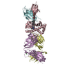
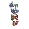
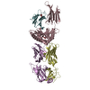
- PDB-4uao: Crystal structure of Apical Membrane Antigen 1 from Plasmodium Kn... -
+ Open data

- Basic information
| Entry | Database: PDB / ID: 4uao | ||||||
|---|---|---|---|---|---|---|---|
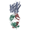
| Title | Crystal structure of Apical Membrane Antigen 1 from Plasmodium Knowlesi in complex with an invasion inhibitory antibody | ||||||
 Components Components |
| ||||||
 Keywords Keywords | CELL INVASION /  Malaria / invasion inhibitory antibody Malaria / invasion inhibitory antibody | ||||||
| Function / homology |  Function and homology information Function and homology informationimmunoglobulin complex, circulating / immunoglobulin receptor binding / complement activation, classical pathway / antigen binding / antibacterial humoral response / membrane / plasma membraneSimilarity search - Function | ||||||
| Biological species |  Plasmodium knowlesi (eukaryote) Plasmodium knowlesi (eukaryote) Rattus norvegicus (Norway rat) Rattus norvegicus (Norway rat) | ||||||
| Method | X-RAY DIFFRACTION / SYNCHROTRON / MOLECULAR REPLACEMENT / Resolution: 3.1 Å | ||||||
 Authors Authors | Vulliez-Le Normand, B. / Saul, F.A. / Bentley, G.A. | ||||||
 Citation Citation | Journal: Plos One / Year: 2015 Title: Crystal Structure of Plasmodium knowlesi Apical Membrane Antigen 1 and Its Complex with an Invasion-Inhibitory Monoclonal Antibody. Authors: Vulliez-Le Normand, B. / Faber, B.W. / Saul, F.A. / van der Eijk, M. / Thomas, A.W. / Singh, B. / Kocken, C.H. / Bentley, G.A. #1: Journal: Clin. Exp. Immunol. / Year: 1982 Title: Rat monoclonal antibodies which inhibit the in vitro multiplication of Plasmodium knowlesi. Authors: Deans, J.A. / Alderson, T. / Thomas, A.W. / Mitchell, G.H. / Lennox, E.S. / Cohen, S. | ||||||
| History |
|
- Structure visualization
| Structure viewer | Molecule: MolmilJmol/JSmol |
|---|
- Downloads & links
-Download
| PDBx/mmCIF format | 4uao.cif.gz | 164.7 KB | Display | PDBx/mmCIF format |
|---|---|---|---|---|
| PDB format | pdb4uao.ent.gz | 127.4 KB | Display | PDB format |
| PDBx/mmJSON format | 4uao.json.gz | Tree view | PDBx/mmJSON format | |
| Others |  Other downloads Other downloads |
-Validation report
| Arichive directory | https://data.pdbj.org/pub/pdb/validation_reports/ua/4uaoftp://data.pdbj.org/pub/pdb/validation_reports/ua/4uao | HTTPS FTP |
|---|
-Related structure data
| Related structure data |  4uv6C  1d5iS  1igfS  1w81S 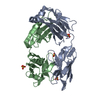 2gcyS C: citing same article ( S: Starting model for refinement |
|---|---|
| Similar structure data |


-Links
 PDBj
PDBj

- Assembly
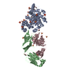
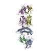
| Deposited unit | 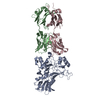
| ||||||||
|---|---|---|---|---|---|---|---|---|---|
| 1 |
| ||||||||
| Unit cell |
|
-Components
| #1: Protein | Mass: 42511.449 Da / Num. of mol.: 1 / Mutation: N107K, S178N, N189E, S240R Source method: isolated from a genetically manipulated source Details: fragment 43-387 of B3L5E1. N-terminal residues Glu 41 and Phe 42 are cloning artifacts. C-terminal residue Gly 388 is a cloning artifact, and following residues Leu 389 to His 410 correspond to expression tags. Source: (gene. exp.) Plasmodium knowlesi (eukaryote) / Gene: PKH_093110 / Plasmid: PPicZalphaA / Production host:  Komagataella pastoris (fungus) / Strain (production host): KM71H / References: UniProt: B3L5E1, UniProt: A0A384KGX8*PLUS Komagataella pastoris (fungus) / Strain (production host): KM71H / References: UniProt: B3L5E1, UniProt: A0A384KGX8*PLUS |
|---|---|
| #2: Antibody | Mass: 23671.410 Da / Num. of mol.: 1 / Source method: isolated from a natural source Details: The sequence for chain B has been deposited to GenBank (KM225619) Source: (natural) Rattus norvegicus (Norway rat) / References: UniProt: P01835*PLUS |
| #3: Antibody | Mass: 23967.773 Da / Num. of mol.: 1 / Source method: isolated from a natural source Details: The sequence for chain C has been deposited to GenBank (KM225620) Source: (natural) Rattus norvegicus (Norway rat) / References: UniProt: P20760*PLUS |
| Compound details | The Fab light and heavy chains are numbered according to the Kabat convention |
-Experimental details
-Experiment
| Experiment | Method: X-RAY DIFFRACTION |
|---|
- Sample preparation
| Crystal | Density Matthews: 2.7 Å3/Da / Density % sol: 54.5 % |
|---|---|
| Crystal grow | Temperature: 290 K / Method: vapor diffusion, hanging drop / pH: 7.4 / Details: 8% PEG 6000, 40 mM Hepes pH7.4, 80 mM NaCl |
-Data collection
| Diffraction | Mean temperature: 100 K |
|---|---|
| Diffraction source | Source: SYNCHROTRON / Site: SOLEIL  / Beamline: PROXIMA 1 / Wavelength: 0.98 Å / Beamline: PROXIMA 1 / Wavelength: 0.98 Å |
| Detector | Type: ADSC QUANTUM 315r / Detector: CCD / Date: Sep 25, 2009 |
| Radiation | Protocol: SINGLE WAVELENGTH / Monochromatic (M) / Laue (L): M / Scattering type: x-ray |
| Radiation wavelength | Wavelength: 0.98 Å / Relative weight: 1 |
| Reflection | Resolution: 3.1→43.35 Å / Num. obs: 17127 / % possible obs: 97.8 % / Redundancy: 3.1 % / Biso Wilson estimate: 71.32 Å2 / Rmerge(I) obs: 0.152 / Net I/σ(I): 7.8 |
| Reflection shell | Resolution: 3.1→3.27 Å / Redundancy: 3.2 % / Rmerge(I) obs: 0.778 / Mean I/σ(I) obs: 1.3 / % possible all: 99.1 |
- Processing
| Software |
| ||||||||||||||||||||||||||||||||||||||||||||||||||||||||||||||||||||||||||||||||||||||||||||||||||||||||||||||||||
|---|---|---|---|---|---|---|---|---|---|---|---|---|---|---|---|---|---|---|---|---|---|---|---|---|---|---|---|---|---|---|---|---|---|---|---|---|---|---|---|---|---|---|---|---|---|---|---|---|---|---|---|---|---|---|---|---|---|---|---|---|---|---|---|---|---|---|---|---|---|---|---|---|---|---|---|---|---|---|---|---|---|---|---|---|---|---|---|---|---|---|---|---|---|---|---|---|---|---|---|---|---|---|---|---|---|---|---|---|---|---|---|---|---|---|---|
| Refinement | Method to determine structure: MOLECULAR REPLACEMENT Starting model: 1W81, 2GCY, 1D5I, 1IGF Resolution: 3.1→40.77 Å / Cor.coef. Fo:Fc: 0.8811 / Cor.coef. Fo:Fc free: 0.8245 / Cross valid method: THROUGHOUT / σ(F): 0 / SU Rfree Blow DPI: 0.475
| ||||||||||||||||||||||||||||||||||||||||||||||||||||||||||||||||||||||||||||||||||||||||||||||||||||||||||||||||||
| Displacement parameters | Biso mean: 70.7 Å2
| ||||||||||||||||||||||||||||||||||||||||||||||||||||||||||||||||||||||||||||||||||||||||||||||||||||||||||||||||||
| Refine analyze | Luzzati coordinate error obs: 0.511 Å | ||||||||||||||||||||||||||||||||||||||||||||||||||||||||||||||||||||||||||||||||||||||||||||||||||||||||||||||||||
| Refinement step | Cycle: 1 / Resolution: 3.1→40.77 Å
| ||||||||||||||||||||||||||||||||||||||||||||||||||||||||||||||||||||||||||||||||||||||||||||||||||||||||||||||||||
| Refine LS restraints |
| ||||||||||||||||||||||||||||||||||||||||||||||||||||||||||||||||||||||||||||||||||||||||||||||||||||||||||||||||||
| LS refinement shell | Resolution: 3.1→3.29 Å / Total num. of bins used: 9
|
