 Movie
Movie Controller

[English] 日本語
 Yorodumi
- PDB-4u89: 4'-phosphopantetheinyl transferase PptT from Mycobacterium tuberc... -
+ Open data

- Basic information
| Entry | Database: PDB / ID: 4u89 | ||||||
|---|---|---|---|---|---|---|---|
| Title | 4'-phosphopantetheinyl transferase PptT from Mycobacterium tuberculosis | ||||||
 Components Components | Phosphopantetheinyl transferase PptT | ||||||
 Keywords Keywords |  TRANSFERASE TRANSFERASE | ||||||
| Function / homology |  Function and homology information Function and homology informationsiderophore metabolic process / enterobactin synthetase complex / holo-[acyl-carrier-protein] synthase / enterobactin biosynthetic process / siderophore biosynthetic process / holo-[acyl-carrier-protein] synthase activity / magnesium ion binding / plasma membraneSimilarity search - Function | ||||||
| Biological species |   Mycobacterium tuberculosis (bacteria) Mycobacterium tuberculosis (bacteria) | ||||||
| Method | X-RAY DIFFRACTION / SYNCHROTRON / SAD / Resolution: 1.4 Å | ||||||
 Authors Authors | Faille, A. / Mourey, L. / Pedelacq, J.D. | ||||||
 Citation Citation | Journal: to be published Title: X-ray structure of the 4'-phosphopantetheinyl transferase PptT from Mycobacterium tuberculosis Authors: Faille, A. / Gavalda, S. / Rottier, K. / Mourey, L. / Pedelacq, J.D. | ||||||
| History |
|
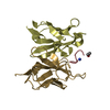
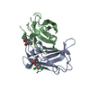
- Structure visualization
| Structure viewer | Molecule: MolmilJmol/JSmol |
|---|
- Downloads & links
-Download
| PDBx/mmCIF format | 4u89.cif.gz | 126.2 KB | Display | PDBx/mmCIF format |
|---|---|---|---|---|
| PDB format | pdb4u89.ent.gz | 95 KB | Display | PDB format |
| PDBx/mmJSON format | 4u89.json.gz | Tree view | PDBx/mmJSON format | |
| Others |  Other downloads Other downloads |
-Validation report
| Arichive directory | https://data.pdbj.org/pub/pdb/validation_reports/u8/4u89ftp://data.pdbj.org/pub/pdb/validation_reports/u8/4u89 | HTTPS FTP |
|---|
-Related structure data
| Similar structure data |
|---|


-Links
 PDBj
PDBj


- Assembly
| Deposited unit | 
| ||||||||
|---|---|---|---|---|---|---|---|---|---|
| 1 |
| ||||||||
| Unit cell |
|
-Components
-Protein , 1 types, 1 molecules A
| #1: Protein | Mass: 27771.666 Da / Num. of mol.: 1 Source method: isolated from a genetically manipulated source Source: (gene. exp.) Mycobacterium tuberculosis (bacteria) / Strain: ATCC 25618 / H37Rv / Gene: pptT, Rv2794c, P425_02911, RVBD_2794c / Plasmid: pTet / Production host: Escherichia coli BL21(DE3) (bacteria) / References: UniProt: O33336 |
|---|
-Non-polymers , 6 types, 286 molecules 


| #2: Chemical | ChemComp-COA / Coenzyme A Mass: 767.534 Da / Num. of mol.: 1 / Source method: obtained synthetically / Formula: C21H36N7O16P3S Mass: 767.534 Da / Num. of mol.: 1 / Source method: obtained synthetically / Formula: C21H36N7O16P3S | ||||||||
|---|---|---|---|---|---|---|---|---|---|
| #3: Chemical | ChemComp-NA /  Mass: 22.990 Da / Num. of mol.: 11 / Source method: obtained synthetically / Formula: Na Mass: 22.990 Da / Num. of mol.: 11 / Source method: obtained synthetically / Formula: Na#4: Chemical | ChemComp-PO4 / | Phosphate Mass: 94.971 Da / Num. of mol.: 1 / Source method: obtained synthetically / Formula: PO4 Mass: 94.971 Da / Num. of mol.: 1 / Source method: obtained synthetically / Formula: PO4#5: Chemical | ChemComp-PG4 / | Polyethylene glycol Mass: 194.226 Da / Num. of mol.: 1 / Source method: obtained synthetically / Formula: C8H18O5 / Comment: precipitant*YM Mass: 194.226 Da / Num. of mol.: 1 / Source method: obtained synthetically / Formula: C8H18O5 / Comment: precipitant*YM#6: Chemical | ChemComp-IMD / | Imidazole Mass: 69.085 Da / Num. of mol.: 1 / Source method: obtained synthetically / Formula: C3H5N2 Mass: 69.085 Da / Num. of mol.: 1 / Source method: obtained synthetically / Formula: C3H5N2#7: Water | ChemComp-HOH / | WaterMass: 18.015 Da / Num. of mol.: 271 / Source method: isolated from a natural source / Formula: H2O |
-Experimental details
-Experiment
| Experiment | Method: X-RAY DIFFRACTION / Number of used crystals: 1 |
|---|
- Sample preparation
| Crystal | Density Matthews: 2.67 Å3/Da / Density % sol: 53.9 % / Description: platelets |
|---|---|
| Crystal grow | Temperature: 285 K / Method: vapor diffusion, hanging drop / pH: 4 Details: 22% PEG 1000, 0.2 M LiSO4, 0.1 M phosphate-citrate pH 4.0 |
-Data collection
| Diffraction |
| |||||||||||||||||||||||||||||||||||||||||||||||||||||||||||||||||||||||||||||||||||||||||||||||||||||||||
|---|---|---|---|---|---|---|---|---|---|---|---|---|---|---|---|---|---|---|---|---|---|---|---|---|---|---|---|---|---|---|---|---|---|---|---|---|---|---|---|---|---|---|---|---|---|---|---|---|---|---|---|---|---|---|---|---|---|---|---|---|---|---|---|---|---|---|---|---|---|---|---|---|---|---|---|---|---|---|---|---|---|---|---|---|---|---|---|---|---|---|---|---|---|---|---|---|---|---|---|---|---|---|---|---|---|---|
| Diffraction source |
| |||||||||||||||||||||||||||||||||||||||||||||||||||||||||||||||||||||||||||||||||||||||||||||||||||||||||
| Detector |
| |||||||||||||||||||||||||||||||||||||||||||||||||||||||||||||||||||||||||||||||||||||||||||||||||||||||||
| Radiation |
| |||||||||||||||||||||||||||||||||||||||||||||||||||||||||||||||||||||||||||||||||||||||||||||||||||||||||
| Radiation wavelength |
| |||||||||||||||||||||||||||||||||||||||||||||||||||||||||||||||||||||||||||||||||||||||||||||||||||||||||
| Reflection | Number: 685637 / Rmerge(I) obs: 0.097 / Χ2: 0.88 / D res high: 2 Å / Num. obs: 32126 / % possible obs: 82.7 | |||||||||||||||||||||||||||||||||||||||||||||||||||||||||||||||||||||||||||||||||||||||||||||||||||||||||
| Diffraction reflection shell |
| |||||||||||||||||||||||||||||||||||||||||||||||||||||||||||||||||||||||||||||||||||||||||||||||||||||||||
| Reflection | Resolution: 1.4→30.316 Å / Num. obs: 58569 / % possible obs: 99.7 % / Observed criterion σ(I): -3 / Redundancy: 6.98 % / Biso Wilson estimate: 21.864 Å2 / Rmerge F obs: 0.998 / Rmerge(I) obs: 0.069 / Rrim(I) all: 0.074 / Χ2: 1.011 / Net I/σ(I): 15.12 / Num. measured all: 408859 | |||||||||||||||||||||||||||||||||||||||||||||||||||||||||||||||||||||||||||||||||||||||||||||||||||||||||
| Reflection shell | Diffraction-ID: 1 / Rejects: 0
|

-Phasing
| Phasing | Method: SAD |
|---|
- Processing
| Software |
| ||||||||||||||||||||||||||||||||||||||||||||||||||||||||||||
|---|---|---|---|---|---|---|---|---|---|---|---|---|---|---|---|---|---|---|---|---|---|---|---|---|---|---|---|---|---|---|---|---|---|---|---|---|---|---|---|---|---|---|---|---|---|---|---|---|---|---|---|---|---|---|---|---|---|---|---|---|---|
| Refinement | Method to determine structure: SAD / Resolution: 1.4→30.32 Å / Cor.coef. Fo:Fc: 0.977 / Cor.coef. Fo:Fc free: 0.971 / WRfactor Rfree: 0.1611 / WRfactor Rwork: 0.1364 / FOM work R set: 0.9134 / SU B: 1.538 / SU ML: 0.028 / SU R Cruickshank DPI: 0.0485 / SU Rfree: 0.0469 / Cross valid method: THROUGHOUT / σ(F): 0 / ESU R: 0.049 / ESU R Free: 0.047 / Stereochemistry target values: MAXIMUM LIKELIHOOD Details: HYDROGENS HAVE BEEN USED IF PRESENT IN THE INPUT U VALUES : REFINED INDIVIDUALLY
| ||||||||||||||||||||||||||||||||||||||||||||||||||||||||||||
| Solvent computation | Ion probe radii: 0.8 Å / Shrinkage radii: 0.8 Å / VDW probe radii: 1.2 Å / Solvent model: MASK | ||||||||||||||||||||||||||||||||||||||||||||||||||||||||||||
| Displacement parameters | Biso max: 109.09 Å2 / Biso mean: 20.226 Å2 / Biso min: 8.26 Å2
| ||||||||||||||||||||||||||||||||||||||||||||||||||||||||||||
| Refinement step | Cycle: final / Resolution: 1.4→30.32 Å
| ||||||||||||||||||||||||||||||||||||||||||||||||||||||||||||
| Refine LS restraints |
| ||||||||||||||||||||||||||||||||||||||||||||||||||||||||||||
| LS refinement shell | Resolution: 1.4→1.436 Å / Total num. of bins used: 20
|
