 Movie
Movie Controller


+ Open data

- Basic information
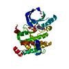
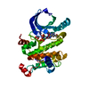
| Entry | Database: PDB / ID: 4u7z | ||||||
|---|---|---|---|---|---|---|---|
| Title | Mitogen-Activated Protein Kinase Kinase (MEK1) bound to G805 | ||||||
 Components Components | Dual specificity mitogen-activated protein kinase kinase 1 | ||||||
 Keywords Keywords | TRANSFERASE/TRANSFERASE Inhibitor /  Kinase / inhibitor / Complex / TRANSFERASE-TRANSFERASE Inhibitor complex Kinase / inhibitor / Complex / TRANSFERASE-TRANSFERASE Inhibitor complex | ||||||
| Function / homology |  Function and homology information Function and homology informationepithelial cell proliferation involved in lung morphogenesis / positive regulation of endodermal cell differentiation / placenta blood vessel development / regulation of axon regeneration / mitogen-activated protein kinase kinase / labyrinthine layer development / type B pancreatic cell proliferation / MAP-kinase scaffold activity / cerebellar cortex formation / Signaling by MAP2K mutants ...epithelial cell proliferation involved in lung morphogenesis / positive regulation of endodermal cell differentiation / placenta blood vessel development / regulation of axon regeneration / mitogen-activated protein kinase kinase / labyrinthine layer development / type B pancreatic cell proliferation / MAP-kinase scaffold activity / cerebellar cortex formation / Signaling by MAP2K mutants / regulation of Golgi inheritance / trachea formation / Negative feedback regulation of MAPK pathway / regulation of early endosome to late endosome transport / positive regulation of axonogenesis / regulation of stress-activated MAPK cascade / Frs2-mediated activation / ERBB2-ERBB3 signaling pathway / protein kinase activator activity / endodermal cell differentiation / face development / MAPK3 (ERK1) activation / Bergmann glial cell differentiation / MAP kinase kinase activity / thyroid gland development / Uptake and function of anthrax toxins / Schwann cell development / keratinocyte differentiation / ERK1 and ERK2 cascade / myelination / protein serine/threonine/tyrosine kinase activity / protein serine/threonine kinase activator activity / MAP3K8 (TPL2)-dependent MAPK1/3 activation / insulin-like growth factor receptor signaling pathway / thymus development / Signal transduction by L1 / cell motility / RAF activation / Signaling by high-kinase activity BRAF mutants / MAP2K and MAPK activation / neuron differentiation / positive regulation of protein serine/threonine kinase activity / Signaling by RAF1 mutants / Signaling by moderate kinase activity BRAF mutants / Paradoxical activation of RAF signaling by kinase inactive BRAF / Signaling downstream of RAS mutants / chemotaxis / MAPK cascade / cellular senescence / Signaling by BRAF and RAF1 fusions / late endosome / heart development / scaffold protein binding / protein tyrosine kinase activity / early endosome / positive regulation of ERK1 and ERK2 cascade / protein kinase activity / negative regulation of cell population proliferation / protein phosphorylation / protein serine kinase activity / focal adhesion / protein serine/threonine kinase activity / centrosome / positive regulation of gene expression / positive regulation of DNA-templated transcription / Golgi apparatus / endoplasmic reticulum / signal transduction / mitochondrion / ATP binding / nucleus / plasma membrane / cytosolSimilarity search - Function | ||||||
| Biological species |  Homo sapiens (human) Homo sapiens (human) | ||||||
| Method | X-RAY DIFFRACTION / SYNCHROTRON / MOLECULAR REPLACEMENT / Resolution: 2.805 Å | ||||||
 Authors Authors | Robarge, K.D. / Ultsch, M.H. / Wiesmann, C. | ||||||
 Citation Citation | Journal: Bioorg.Med.Chem.Lett. / Year: 2014 Title: Structure based design of novel 6,5 heterobicyclic mitogen-activated protein kinase kinase (MEK) inhibitors leading to the discovery of imidazo[1,5-a] pyrazine G-479. Authors: Robarge, K.D. / Lee, W. / Eigenbrot, C. / Ultsch, M. / Wiesmann, C. / Heald, R. / Price, S. / Hewitt, J. / Jackson, P. / Savy, P. / Burton, B. / Choo, E.F. / Pang, J. / Boggs, J. / Yang, A. ...Authors: Robarge, K.D. / Lee, W. / Eigenbrot, C. / Ultsch, M. / Wiesmann, C. / Heald, R. / Price, S. / Hewitt, J. / Jackson, P. / Savy, P. / Burton, B. / Choo, E.F. / Pang, J. / Boggs, J. / Yang, A. / Yang, X. / Baumgardner, M. | ||||||
| History |
|
- Structure visualization
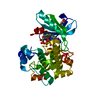
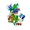
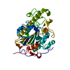
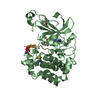
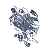
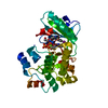
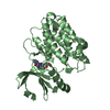
| Structure viewer | Molecule: MolmilJmol/JSmol |
|---|
- Downloads & links
-Download
| PDBx/mmCIF format | 4u7z.cif.gz | 136.6 KB | Display | PDBx/mmCIF format |
|---|---|---|---|---|
| PDB format | pdb4u7z.ent.gz | 103.9 KB | Display | PDB format |
| PDBx/mmJSON format | 4u7z.json.gz | Tree view | PDBx/mmJSON format | |
| Others |  Other downloads Other downloads |
-Validation report
| Arichive directory | https://data.pdbj.org/pub/pdb/validation_reports/u7/4u7zftp://data.pdbj.org/pub/pdb/validation_reports/u7/4u7z | HTTPS FTP |
|---|
-Related structure data
| Related structure data |  4u80C 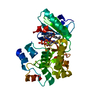 4u81C 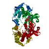 1s9jS S: Starting model for refinement C: citing same article ( |
|---|---|
| Similar structure data |


-Links
 PDBj
PDBj


- Assembly
| Deposited unit | 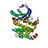
| ||||||||
|---|---|---|---|---|---|---|---|---|---|
| 1 |
| ||||||||
| Unit cell |
|
-Components
| #1: Protein | Mass: 37929.625 Da / Num. of mol.: 1 / Fragment: kinase domain (UNP residues 62-393) Source method: isolated from a genetically manipulated source Source: (gene. exp.) Homo sapiens (human) / Gene: MAP2K1, MEK1, PRKMK1 / Plasmid: pET24B / Production host:  Escherichia coli (E. coli) / Strain (production host): Rosetta 2 PLysS Escherichia coli (E. coli) / Strain (production host): Rosetta 2 PLysSReferences: UniProt: Q02750, mitogen-activated protein kinase kinase |
|---|---|
| #2: Chemical | ChemComp-MG /   Mass: 24.305 Da / Num. of mol.: 1 / Source method: obtained synthetically / Formula: Mg Mass: 24.305 Da / Num. of mol.: 1 / Source method: obtained synthetically / Formula: Mg |
| #3: Chemical | ChemComp-3EW / Selumetinib  Mass: 457.681 Da / Num. of mol.: 1 / Source method: obtained synthetically / Formula: C17H15BrClFN4O3 / Comment: medication*YM Mass: 457.681 Da / Num. of mol.: 1 / Source method: obtained synthetically / Formula: C17H15BrClFN4O3 / Comment: medication*YM |
| #4: Chemical | ChemComp-ANP /   Mass: 506.196 Da / Num. of mol.: 1 / Source method: obtained synthetically / Formula: C10H17N6O12P3 / Comment: AMP-PNP, energy-carrying molecule analogue*YM Mass: 506.196 Da / Num. of mol.: 1 / Source method: obtained synthetically / Formula: C10H17N6O12P3 / Comment: AMP-PNP, energy-carrying molecule analogue*YM |
| #5: Water | ChemComp-HOH / Water Mass: 18.015 Da / Num. of mol.: 11 / Source method: isolated from a natural source / Formula: H2O Mass: 18.015 Da / Num. of mol.: 11 / Source method: isolated from a natural source / Formula: H2O |
-Experimental details
-Experiment
| Experiment | Method: X-RAY DIFFRACTION / Number of used crystals: 1 |
|---|
- Sample preparation
| Crystal | Density Matthews: 3.3 Å3/Da / Density % sol: 62.77 % |
|---|---|
| Crystal grow | Temperature: 291 K / Method: vapor diffusion, hanging drop / pH: 6.9 Details: The protein was concentrated to 15mg/ml and incubated with 10 fold molar excess inhibitor plus 1mM MgAMP-PNP before crystallization. MEK1 crystals grew from hanging drop vapor diffusion ...Details: The protein was concentrated to 15mg/ml and incubated with 10 fold molar excess inhibitor plus 1mM MgAMP-PNP before crystallization. MEK1 crystals grew from hanging drop vapor diffusion using 12% w/v PEG 8000, 0.4M NH4H2PO4 and 0.1M HEPES pH 6.9 at 18 degC. PH range: 6.9 |
-Data collection
| Diffraction | Mean temperature: 93 K |
|---|---|
| Diffraction source | Source: SYNCHROTRON / Site: SSRL  / Beamline: BL11-1 / Wavelength: 0.987 Å / Beamline: BL11-1 / Wavelength: 0.987 Å |
| Detector | Type: DECTRIS PILATUS 6M / Detector: PIXEL / Date: Nov 6, 2006 |
| Radiation | Monochromator: Side scattering bent cube-root I-beam single crystal; asymmetric cut 4.965 degs Protocol: SINGLE WAVELENGTH / Monochromatic (M) / Laue (L): M / Scattering type: x-ray |
| Radiation wavelength | Wavelength: 0.987 Å / Relative weight: 1 |
| Reflection | Resolution: 2.7→50 Å / Num. all: 12041 / Num. obs: 12041 / % possible obs: 80.6 % / Observed criterion σ(I): 2 / Redundancy: 1.8 % / Biso Wilson estimate: 76.3 Å2 / Rsym value: 0.08 / Net I/σ(I): 17.9 |
| Reflection shell | Resolution: 2.7→2.8 Å / Redundancy: 1.8 % / Rmerge(I) obs: 0.588 / Mean I/σ(I) obs: 1.2 / % possible all: 88.6 |
- Processing
| Software | Name: PHENIX / Version: (phenix.refine: 1.8.2_1309) / Classification: refinement | |||||||||||||||||||||||||||||||||||||||||||||||||||||||||||||||||||||||||||||||||||||||||||||||||||||||||||||||||||||||||||||
|---|---|---|---|---|---|---|---|---|---|---|---|---|---|---|---|---|---|---|---|---|---|---|---|---|---|---|---|---|---|---|---|---|---|---|---|---|---|---|---|---|---|---|---|---|---|---|---|---|---|---|---|---|---|---|---|---|---|---|---|---|---|---|---|---|---|---|---|---|---|---|---|---|---|---|---|---|---|---|---|---|---|---|---|---|---|---|---|---|---|---|---|---|---|---|---|---|---|---|---|---|---|---|---|---|---|---|---|---|---|---|---|---|---|---|---|---|---|---|---|---|---|---|---|---|---|---|
| Refinement | Method to determine structure: MOLECULAR REPLACEMENT Starting model: 1S9J Resolution: 2.805→19.659 Å / SU ML: 0.38 / Cross valid method: FREE R-VALUE / σ(F): 1.39 / Phase error: 23.39 / Stereochemistry target values: ML
| |||||||||||||||||||||||||||||||||||||||||||||||||||||||||||||||||||||||||||||||||||||||||||||||||||||||||||||||||||||||||||||
| Solvent computation | Shrinkage radii: 0.9 Å / VDW probe radii: 1.11 Å / Solvent model: FLAT BULK SOLVENT MODEL | |||||||||||||||||||||||||||||||||||||||||||||||||||||||||||||||||||||||||||||||||||||||||||||||||||||||||||||||||||||||||||||
| Refinement step | Cycle: LAST / Resolution: 2.805→19.659 Å
| |||||||||||||||||||||||||||||||||||||||||||||||||||||||||||||||||||||||||||||||||||||||||||||||||||||||||||||||||||||||||||||
| Refine LS restraints |
| |||||||||||||||||||||||||||||||||||||||||||||||||||||||||||||||||||||||||||||||||||||||||||||||||||||||||||||||||||||||||||||
| LS refinement shell |
| |||||||||||||||||||||||||||||||||||||||||||||||||||||||||||||||||||||||||||||||||||||||||||||||||||||||||||||||||||||||||||||
| Refinement TLS params. | Method: refined / Refine-ID: X-RAY DIFFRACTION
| |||||||||||||||||||||||||||||||||||||||||||||||||||||||||||||||||||||||||||||||||||||||||||||||||||||||||||||||||||||||||||||
| Refinement TLS group |
|
