 Movie
Movie Controller

[English] 日本語
 Yorodumi
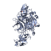
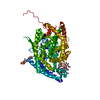
- PDB-4u4p: Crystal structure of the human condensin SMC hinge domain heterod... -
+ Open data

- Basic information
| Entry | Database: PDB / ID: 4u4p | ||||||
|---|---|---|---|---|---|---|---|
| Title | Crystal structure of the human condensin SMC hinge domain heterodimer with short coiled coils | ||||||
 Components Components |
| ||||||
 Keywords Keywords |  PROTEIN BINDING / Condensin / SMC PROTEIN BINDING / Condensin / SMC | ||||||
| Function / homology |  Function and homology information Function and homology informationcondensin complex => GO:0000796 / condensin complex => GO:0000796 / meiotic chromosome condensation / condensin complex / kinetochore organization / meiotic chromosome segregation / mitotic chromosome condensation / Condensation of Prometaphase Chromosomes / nuclear chromosome / mitotic sister chromatid segregation ...condensin complex => GO:0000796 / condensin complex => GO:0000796 / meiotic chromosome condensation / condensin complex / kinetochore organization / meiotic chromosome segregation / mitotic chromosome condensation / Condensation of Prometaphase Chromosomes / nuclear chromosome / mitotic sister chromatid segregation / chromosome, centromeric region / condensed chromosome / Condensation of Prophase Chromosomes / single-stranded DNA binding / nuclear speck / cell division / nucleolus / extracellular exosome / nucleoplasm / ATP binding / nucleus / cytosol / cytoplasmSimilarity search - Function | ||||||
| Biological species |  Homo sapiens (human) Homo sapiens (human) | ||||||
| Method | X-RAY DIFFRACTION / SYNCHROTRON / MOLECULAR REPLACEMENT / molecular replacement / Resolution: 1.89 Å | ||||||
 Authors Authors | Uchiyama, S. / Kawahara, K. / Hosokawa, Y. / Fukakusa, S. / Oki, H. / Nakamura, S. / Noda, M. / Takino, R. / Miyahara, Y. / Maruno, T. ...Uchiyama, S. / Kawahara, K. / Hosokawa, Y. / Fukakusa, S. / Oki, H. / Nakamura, S. / Noda, M. / Takino, R. / Miyahara, Y. / Maruno, T. / Kobayashi, Y. / Ohkubo, T. / Fukui, K. | ||||||
 Citation Citation | Journal: to be published Title: Structural basis for dimer information and DNA recognition of human SMC proteins Authors: Uchiyama, S. / Kawahara, K. / Hosokawa, Y. / Fukakusa, S. / Oki, H. / Nakamura, S. / Noda, M. / Takino, R. / Miyahara, Y. / Maruno, T. / Kobayashi, Y. / Ohkubo, T. / Fukui, K. | ||||||
| History |
|
- Structure visualization
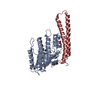
| Structure viewer | Molecule: MolmilJmol/JSmol |
|---|
- Downloads & links
-Download
| PDBx/mmCIF format | 4u4p.cif.gz | 105.4 KB | Display | PDBx/mmCIF format |
|---|---|---|---|---|
| PDB format | pdb4u4p.ent.gz | 77.8 KB | Display | PDB format |
| PDBx/mmJSON format | 4u4p.json.gz | Tree view | PDBx/mmJSON format | |
| Others |  Other downloads Other downloads |
-Validation report
| Arichive directory | https://data.pdbj.org/pub/pdb/validation_reports/u4/4u4pftp://data.pdbj.org/pub/pdb/validation_reports/u4/4u4p | HTTPS FTP |
|---|
-Related structure data
| Related structure data |  3l51S S: Starting model for refinement |
|---|---|
| Similar structure data |


-Links
 PDBj
PDBj


- Assembly
| Deposited unit | 
| ||||||||
|---|---|---|---|---|---|---|---|---|---|
| 1 |
| ||||||||
| Unit cell |
|
-Components
| #1: Protein | Mass: 26278.309 Da / Num. of mol.: 1 / Fragment: UNP RESIDUES 476-707 Source method: isolated from a genetically manipulated source Source: (gene. exp.) Homo sapiens (human) / Gene: SMC2, CAPE, SMC2L1, PRO0324 / Production host:  Escherichia coli (E. coli) / References: UniProt: O95347 Escherichia coli (E. coli) / References: UniProt: O95347 |
|---|---|
| #2: Protein | Mass: 27906.041 Da / Num. of mol.: 1 / Fragment: UNP RESIDUES 566-809 Source method: isolated from a genetically manipulated source Source: (gene. exp.) Homo sapiens (human) / Gene: SMC4, CAPC, SMC4L1 / Production host: Escherichia coli (E. coli) / References: UniProt: Q9NTJ3 |
| #3: Water | ChemComp-HOH / Water Mass: 18.015 Da / Num. of mol.: 303 / Source method: isolated from a natural source / Formula: H2O Mass: 18.015 Da / Num. of mol.: 303 / Source method: isolated from a natural source / Formula: H2O |
-Experimental details
-Experiment
| Experiment | Method: X-RAY DIFFRACTION / Number of used crystals: 1 |
|---|
- Sample preparation
| Crystal | Density Matthews: 2.23 Å3/Da / Density % sol: 44.86 % |
|---|---|
| Crystal grow | Temperature: 277 K / Method: vapor diffusion, sitting drop / pH: 8.5 / Details: 0.1M Tris-HCl pH 8.5, 19% PEG 3350, 50mM NaCl |
-Data collection
| Diffraction | Mean temperature: 100 K | |||||||||||||||||||||||||||
|---|---|---|---|---|---|---|---|---|---|---|---|---|---|---|---|---|---|---|---|---|---|---|---|---|---|---|---|---|
| Diffraction source | Source: SYNCHROTRON / Site: Photon Factory  / Beamline: BL-17A / Wavelength: 0.98 Å / Beamline: BL-17A / Wavelength: 0.98 Å | |||||||||||||||||||||||||||
| Detector | Type: ADSC QUANTUM 315r / Detector: CCD / Date: Oct 9, 2011 | |||||||||||||||||||||||||||
| Radiation | Protocol: SINGLE WAVELENGTH / Monochromatic (M) / Laue (L): M / Scattering type: x-ray | |||||||||||||||||||||||||||
| Radiation wavelength | Wavelength: 0.98 Å / Relative weight: 1 | |||||||||||||||||||||||||||
| Reflection | Resolution: 1.89→46.99 Å / Num. obs: 47639 / % possible obs: 98.4 % / Redundancy: 4.4 % / Biso Wilson estimate: 23.43 Å2 / CC1/2: 0.997 / Rmerge(I) obs: 0.066 / Rpim(I) all: 0.044 / Net I/σ(I): 10.8 / Num. measured all: 209398 / Scaling rejects: 29 | |||||||||||||||||||||||||||
| Reflection shell | Diffraction-ID: 1 / Rejects: 0
|
-Phasing
| Phasing | Method: molecular replacement |
|---|
- Processing
| Software | Name: PHENIX / Version: 1.9_1692 / Classification: refinement | |||||||||||||||||||||||||||||||||||||||||||||||||||||||||||||||||||||||||||||||||||||||||||||||||||||||||
|---|---|---|---|---|---|---|---|---|---|---|---|---|---|---|---|---|---|---|---|---|---|---|---|---|---|---|---|---|---|---|---|---|---|---|---|---|---|---|---|---|---|---|---|---|---|---|---|---|---|---|---|---|---|---|---|---|---|---|---|---|---|---|---|---|---|---|---|---|---|---|---|---|---|---|---|---|---|---|---|---|---|---|---|---|---|---|---|---|---|---|---|---|---|---|---|---|---|---|---|---|---|---|---|---|---|---|
| Refinement | Method to determine structure: MOLECULAR REPLACEMENT Starting model: 3L51 Resolution: 1.89→45.191 Å / SU ML: 0.21 / Cross valid method: FREE R-VALUE / Phase error: 22.29 / Stereochemistry target values: ML
| |||||||||||||||||||||||||||||||||||||||||||||||||||||||||||||||||||||||||||||||||||||||||||||||||||||||||
| Solvent computation | Shrinkage radii: 0.9 Å / VDW probe radii: 1.11 Å / Solvent model: FLAT BULK SOLVENT MODEL | |||||||||||||||||||||||||||||||||||||||||||||||||||||||||||||||||||||||||||||||||||||||||||||||||||||||||
| Refinement step | Cycle: LAST / Resolution: 1.89→45.191 Å
| |||||||||||||||||||||||||||||||||||||||||||||||||||||||||||||||||||||||||||||||||||||||||||||||||||||||||
| Refine LS restraints |
| |||||||||||||||||||||||||||||||||||||||||||||||||||||||||||||||||||||||||||||||||||||||||||||||||||||||||
| LS refinement shell |
|
