 Movie
Movie Controller

[English] 日本語
 Yorodumi
- PDB-4u4m: Crystal structure of 0.5M urea unfolded YagE, a KDG aldolase prot... -
+ Open data

- Basic information
| Entry | Database: PDB / ID: 4u4m | |||||||||
|---|---|---|---|---|---|---|---|---|---|---|
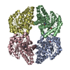
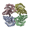
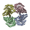
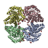
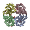
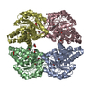
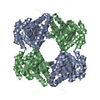
| Title | Crystal structure of 0.5M urea unfolded YagE, a KDG aldolase protein in complex with Pyruvate | |||||||||
 Components Components | YagE Banisteriopsis caapi Banisteriopsis caapi | |||||||||
 Keywords Keywords | SUGAR BINDING PROTEIN / TIM barrel / NAL superfamily / KDGA / Schiff base / catalytic triad / Chemical denaturant / Urea promoted unfolding | |||||||||
| Function / homology |  Function and homology information2-dehydro-3-deoxy-D-pentonate aldolase / aldonic acid catabolic process / 2-dehydro-3-deoxy-D-pentonate aldolase activity / 2-dehydro-3-deoxy-D-gluconate aldolase / 2-dehydro-3-deoxy-D-gluconate aldolase activity / identical protein binding / cytosol Function and homology information2-dehydro-3-deoxy-D-pentonate aldolase / aldonic acid catabolic process / 2-dehydro-3-deoxy-D-pentonate aldolase activity / 2-dehydro-3-deoxy-D-gluconate aldolase / 2-dehydro-3-deoxy-D-gluconate aldolase activity / identical protein binding / cytosolSimilarity search - Function | |||||||||
| Biological species |  Escherichia coli K-12 (bacteria) Escherichia coli K-12 (bacteria) | |||||||||
| Method | X-RAY DIFFRACTION / SYNCHROTRON / MOLECULAR REPLACEMENT / Resolution: 3.09 Å | |||||||||
 Authors Authors | Manoj Kumar, P. / Bhaskar, V. / Manicka, S. / Krishnaswamy, S. | |||||||||
 Citation Citation | Journal: To be published Title: Crystal structure of 0.5M urea unfolded YagE, a KDG aldolase protein in complex with Pyruvate Authors: Manoj Kumar, P. / Bhaskar, V. / Manicka, S. / Krishnaswamy, S. #1: Journal: Proteins / Year: 2011Title: Identification of biochemical and putative biological role of a xenolog from Escherichia coli using structural analysis. Authors: Bhaskar, V. / Kumar, M. / Manicka, S. / Tripathi, S. / Venkatraman, A. / Krishnaswamy, S. #2: Journal: Proteins / Year: 2008Title: Crystal structure of YagE, a putative DHDPS-like protein from Escherichia coli K12 Authors: Manicka, S. / Unger, T. / Albeck, S. / Dym, O. / Greenblatt, H.M. / Bourenkov, G. / Lamzin, V. / Krishnaswamy, S. / Sussman, J.L. | |||||||||
| History |
|
- Structure visualization
| Structure viewer | Molecule: MolmilJmol/JSmol |
|---|
- Downloads & links
-Download
| PDBx/mmCIF format | 4u4m.cif.gz | 228.7 KB | Display | PDBx/mmCIF format |
|---|---|---|---|---|
| PDB format | pdb4u4m.ent.gz | 184.3 KB | Display | PDB format |
| PDBx/mmJSON format | 4u4m.json.gz | Tree view | PDBx/mmJSON format | |
| Others |  Other downloads Other downloads |
-Validation report
| Arichive directory | https://data.pdbj.org/pub/pdb/validation_reports/u4/4u4mftp://data.pdbj.org/pub/pdb/validation_reports/u4/4u4m | HTTPS FTP |
|---|
-Related structure data
| Related structure data | 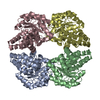 2v8zS S: Starting model for refinement |
|---|---|
| Similar structure data |


-Links
 PDBj
PDBj

- Assembly
| Deposited unit | 
| ||||||||||||||||||||||||||||||||||||||||||||||||||||||||||||||||||||||||||||||||||||||||||||||||||
|---|---|---|---|---|---|---|---|---|---|---|---|---|---|---|---|---|---|---|---|---|---|---|---|---|---|---|---|---|---|---|---|---|---|---|---|---|---|---|---|---|---|---|---|---|---|---|---|---|---|---|---|---|---|---|---|---|---|---|---|---|---|---|---|---|---|---|---|---|---|---|---|---|---|---|---|---|---|---|---|---|---|---|---|---|---|---|---|---|---|---|---|---|---|---|---|---|---|---|---|
| 1 |
| ||||||||||||||||||||||||||||||||||||||||||||||||||||||||||||||||||||||||||||||||||||||||||||||||||
| Unit cell |
| ||||||||||||||||||||||||||||||||||||||||||||||||||||||||||||||||||||||||||||||||||||||||||||||||||
| Noncrystallographic symmetry (NCS) | NCS domain:
NCS domain segments: Component-ID: 0 / Beg auth comp-ID: ALA / Beg label comp-ID: ALA / End auth comp-ID: CYS / End label comp-ID: CYS / Refine code: 0 / Auth seq-ID: 12 - 309 / Label seq-ID: 1 - 298
NCS ensembles :
|
-Components
| #1: Protein | Banisteriopsis caapi Mass: 32119.775 Da / Num. of mol.: 4 Source method: isolated from a genetically manipulated source Source: (gene. exp.) Escherichia coli K-12 (bacteria) / Strain: K12 / Gene: YAGE / Plasmid: pET28A / Details (production host): PET28TEVH/YAGE / Production host: ESCHERICHIA COLI (E. coli) / Strain (production host): BL21 (DE3) / References: UniProt: P75682*PLUS#2: Chemical | ChemComp-URE / Urea  Mass: 60.055 Da / Num. of mol.: 10 / Source method: obtained synthetically / Formula: CH4N2O Mass: 60.055 Da / Num. of mol.: 10 / Source method: obtained synthetically / Formula: CH4N2O#3: Chemical | ChemComp-PYR / Pyruvic acid  Mass: 88.062 Da / Num. of mol.: 4 / Source method: obtained synthetically / Formula: C3H4O3 Mass: 88.062 Da / Num. of mol.: 4 / Source method: obtained synthetically / Formula: C3H4O3#4: Chemical | ChemComp-EDO / Ethylene glycol  Mass: 62.068 Da / Num. of mol.: 10 / Source method: obtained synthetically / Formula: C2H6O2 Mass: 62.068 Da / Num. of mol.: 10 / Source method: obtained synthetically / Formula: C2H6O2#5: Water | ChemComp-HOH / | Water Mass: 18.015 Da / Num. of mol.: 3 / Source method: isolated from a natural source / Formula: H2O Mass: 18.015 Da / Num. of mol.: 3 / Source method: isolated from a natural source / Formula: H2OSequence details | THE SEQUENCE OF THIS PROTEIN WAS NOT AVAILABLE AT THE UNIPROT KNOWLEDGEBASE DATABASE (UNIPROTKB) AT ...THE SEQUENCE OF THIS PROTEIN WAS NOT AVAILABLE AT THE UNIPROT KNOWLEDGEB | |
|---|
-Experimental details
-Experiment
| Experiment | Method: X-RAY DIFFRACTION |
|---|
- Sample preparation
| Crystal | Density Matthews: 2.39 Å3/Da / Density % sol: 47.23 % |
|---|---|
| Crystal grow | Temperature: 298 K / Method: microbatch / pH: 6.5 / Details: 15% PEG3350, 200mM MgCl2, 100mM HEPES pH 6.5 |
-Data collection
| Diffraction | Mean temperature: 100 K |
|---|---|
| Diffraction source | Source: SYNCHROTRON / Site: ESRF  / Beamline: BM14 / Wavelength: 0.9747 Å / Beamline: BM14 / Wavelength: 0.9747 Å |
| Detector | Type: MARMOSAIC 225 mm CCD / Detector: CCD / Date: May 28, 2012 |
| Radiation | Protocol: SINGLE WAVELENGTH / Monochromatic (M) / Laue (L): M / Scattering type: x-ray |
| Radiation wavelength | Wavelength: 0.9747 Å / Relative weight: 1 |
| Reflection | Resolution: 3.09→19.88 Å / Num. obs: 23206 / % possible obs: 99.67 % / Redundancy: 10.9 % / Biso Wilson estimate: 33.36 Å2 / Rmerge(I) obs: 0.2197 / Net I/σ(I): 11.88 |
| Reflection shell | Resolution: 3.09→3.2 Å / Redundancy: 11.2 % / Rmerge(I) obs: 0.6216 / Mean I/σ(I) obs: 5.17 / % possible all: 99.69 |
- Processing
| Software | Name: REFMAC / Version: 5.7.0032 / Classification: refinement | ||||||||||||||||||||||||||||||||||||||||||||||||||||||||||||||||||||||||||||||||||||||||||||||||||||||||||||||||||||||||||||||||||||||||||||||||||||||||||||||||||||||||||||||||||||||
|---|---|---|---|---|---|---|---|---|---|---|---|---|---|---|---|---|---|---|---|---|---|---|---|---|---|---|---|---|---|---|---|---|---|---|---|---|---|---|---|---|---|---|---|---|---|---|---|---|---|---|---|---|---|---|---|---|---|---|---|---|---|---|---|---|---|---|---|---|---|---|---|---|---|---|---|---|---|---|---|---|---|---|---|---|---|---|---|---|---|---|---|---|---|---|---|---|---|---|---|---|---|---|---|---|---|---|---|---|---|---|---|---|---|---|---|---|---|---|---|---|---|---|---|---|---|---|---|---|---|---|---|---|---|---|---|---|---|---|---|---|---|---|---|---|---|---|---|---|---|---|---|---|---|---|---|---|---|---|---|---|---|---|---|---|---|---|---|---|---|---|---|---|---|---|---|---|---|---|---|---|---|---|---|
| Refinement | Method to determine structure: MOLECULAR REPLACEMENT Starting model: 2V8Z Resolution: 3.09→19.88 Å / Cor.coef. Fo:Fc: 0.935 / Cor.coef. Fo:Fc free: 0.916 / SU B: 20.441 / SU ML: 0.36 / Cross valid method: THROUGHOUT / ESU R Free: 0.494 / Stereochemistry target values: MAXIMUM LIKELIHOOD / Details: HYDROGENS HAVE BEEN ADDED IN THE RIDING POSITIONS
| ||||||||||||||||||||||||||||||||||||||||||||||||||||||||||||||||||||||||||||||||||||||||||||||||||||||||||||||||||||||||||||||||||||||||||||||||||||||||||||||||||||||||||||||||||||||
| Solvent computation | Ion probe radii: 0.8 Å / Shrinkage radii: 0.8 Å / VDW probe radii: 1.2 Å / Solvent model: MASK | ||||||||||||||||||||||||||||||||||||||||||||||||||||||||||||||||||||||||||||||||||||||||||||||||||||||||||||||||||||||||||||||||||||||||||||||||||||||||||||||||||||||||||||||||||||||
| Displacement parameters | Biso mean: 24.599 Å2
| ||||||||||||||||||||||||||||||||||||||||||||||||||||||||||||||||||||||||||||||||||||||||||||||||||||||||||||||||||||||||||||||||||||||||||||||||||||||||||||||||||||||||||||||||||||||
| Refinement step | Cycle: 1 / Resolution: 3.09→19.88 Å
| ||||||||||||||||||||||||||||||||||||||||||||||||||||||||||||||||||||||||||||||||||||||||||||||||||||||||||||||||||||||||||||||||||||||||||||||||||||||||||||||||||||||||||||||||||||||
| Refine LS restraints |
|