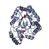
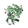
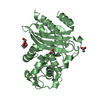
- PDB-4u3v: Crystal structure of the trans-acyltransferase polyketide synthas... -
+
Open data
ID or keywords:
Loading...
-
Basic information
Entry
Database: PDB / ID: 4u3v
Title
Crystal structure of the trans-acyltransferase polyketide synthase enoyl-isomerase
Components
Polyketide synthase PksR
Keywords
ISOMERASE / double-hotdog / polyketide / trans-AT
Function / homology
Function and homology information
biosynthetic process / acyltransferase activity, transferring groups other than amino-acyl groups / hydrolase activity, acting on ester bonds / phosphopantetheine binding / Transferases; Acyltransferases; Transferring groups other than aminoacyl groups / cytoplasm Similarity search - Function
Resolution: 1.73→35.37 Å / Cor.coef. Fo:Fc: 0.957 / Cor.coef. Fo:Fc free: 0.947 / Cross valid method: THROUGHOUT / ESU R: 0.129 / ESU R Free: 0.119 / Stereochemistry target values: MAXIMUM LIKELIHOOD / Details: HYDROGENS HAVE BEEN USED IF PRESENT IN THE INPUT
Rfactor
Num. reflection
% reflection
Selection details
Rfree
0.23855
1380
5.1 %
RANDOM
Rwork
0.21356
-
-
-
obs
0.21478
25889
97.57 %
-
Solvent computation
Ion probe radii: 0.8 Å / Shrinkage radii: 0.8 Å / VDW probe radii: 1.2 Å / Solvent model: MASK
 Movie
Movie Controller

 Yorodumi
 Open data
 Basic information
 Components
 Keywords
 ISOMERASE / double-hotdog /
  Function and homology information
 Authors
 United States, 1items
  Citation
 Structure visualization
 Downloads & links
 Other downloads
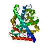


 PDBj
PDBj

 Assembly
 Mass: 18.015 Da / Num. of mol.: 131 / Source method: isolated from a natural source / Formula: H2O
 Sample preparation
 Processing