 Movie
Movie Controller


+ Open data

- Basic information
| Entry | Database: PDB / ID: 4tzf | ||||||
|---|---|---|---|---|---|---|---|
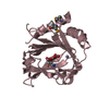
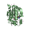
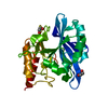
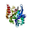
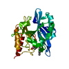
| Title | Structure of metallo-beta lactamase | ||||||
 Components Components | NDM-8 metallo-beta-lactamase | ||||||
 Keywords Keywords |  HYDROLASE / metallo-beta-lactamase / antibiotic resistance HYDROLASE / metallo-beta-lactamase / antibiotic resistance | ||||||
| Function / homology |  Function and homology information Function and homology information | ||||||
| Biological species |  Escherichia coli (E. coli) Escherichia coli (E. coli) | ||||||
| Method | X-RAY DIFFRACTION / SYNCHROTRON / MOLECULAR REPLACEMENT / Resolution: 1.22 Å | ||||||
 Authors Authors | Ferguson, J.A. / Makena, A. / Brem, J. / McDonough, M.A. / Schofield, C.J. | ||||||
 Citation Citation | Journal: To Be Published Title: structure of metallo-beta lactamase Authors: Ferguson, J.A. / Makena, A. / Brem, J. / McDonough, M.A. / Schofield, C.J. | ||||||
| History |
|
- Structure visualization
| Structure viewer | Molecule: MolmilJmol/JSmol |
|---|
- Downloads & links
-Download
| PDBx/mmCIF format | 4tzf.cif.gz | 113.5 KB | Display | PDBx/mmCIF format |
|---|---|---|---|---|
| PDB format | pdb4tzf.ent.gz | 85 KB | Display | PDB format |
| PDBx/mmJSON format | 4tzf.json.gz | Tree view | PDBx/mmJSON format | |
| Others |  Other downloads Other downloads |
-Validation report
| Arichive directory | https://data.pdbj.org/pub/pdb/validation_reports/tz/4tzfftp://data.pdbj.org/pub/pdb/validation_reports/tz/4tzf | HTTPS FTP |
|---|
-Related structure data
| Related structure data |  3spuS S: Starting model for refinement |
|---|---|
| Similar structure data |


-Links
 PDBj
PDBj

- Assembly
| Deposited unit | 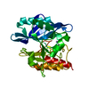
| ||||||||
|---|---|---|---|---|---|---|---|---|---|
| 1 |
| ||||||||
| Unit cell |
|
-Components
| #1: Protein | Mass: 24267.299 Da / Num. of mol.: 1 / Fragment: UNP Residues 42-270 Source method: isolated from a genetically manipulated source Source: (gene. exp.) Escherichia coli (E. coli) / Gene: blaNDM-8 / Plasmid: OPINF / Production host: Escherichia coli BL21(DE3) (bacteria) / Variant (production host): PlysS / References: UniProt: M1VE66 | ||
|---|---|---|---|
| #2: Chemical |   Mass: 65.409 Da / Num. of mol.: 2 / Source method: obtained synthetically / Formula: Zn Mass: 65.409 Da / Num. of mol.: 2 / Source method: obtained synthetically / Formula: Zn#3: Water | ChemComp-HOH / | Water Mass: 18.015 Da / Num. of mol.: 287 / Source method: isolated from a natural source / Formula: H2O Mass: 18.015 Da / Num. of mol.: 287 / Source method: isolated from a natural source / Formula: H2O |
-Experimental details
-Experiment
| Experiment | Method: X-RAY DIFFRACTION |
|---|
- Sample preparation
| Crystal | Density Matthews: 2.09 Å3/Da / Density % sol: 41.09 % / Description: cubic |
|---|---|
| Crystal grow | Temperature: 298 K / Method: vapor diffusion, sitting drop / pH: 5.5 / Details: 0.1 M Bis Tris pH 5.5, 25% PEG 3350 |
-Data collection
| Diffraction | Mean temperature: 100 K |
|---|---|
| Diffraction source | Source: SYNCHROTRON / Site: Diamond  / Beamline: I02 / Wavelength: 0.9793 Å / Beamline: I02 / Wavelength: 0.9793 Å |
| Detector | Type: DECTRIS PILATUS 6M-F / Detector: PIXEL / Date: Feb 10, 2014 |
| Radiation | Monochromator: double crystal / Protocol: SINGLE WAVELENGTH / Monochromatic (M) / Laue (L): M / Scattering type: x-ray |
| Radiation wavelength | Wavelength: 0.9793 Å / Relative weight: 1 |
| Reflection | Resolution: 1.22→42.47 Å / Num. all: 61432 / Num. obs: 61432 / % possible obs: 99.7 % / Redundancy: 6.2 % / Rmerge(I) obs: 0.065 / Net I/av σ(I): 13.4 / Net I/σ(I): 13.4 |
| Reflection shell | Resolution: 1.22→1.25 Å / Redundancy: 6.1 % / Rmerge(I) obs: 0.66 / Mean I/σ(I) obs: 2.4 / % possible all: 99.6 |
- Processing
| Software |
| ||||||||||||||||||||||||||||||||||||||||||||||||||||||||||||||||||||||||||||||||||||||||||||||||||||||||||||||||||||||||||||||||||||||||||||||||||||||||||||||||||||||||||||||||||||||||||||||||||||
|---|---|---|---|---|---|---|---|---|---|---|---|---|---|---|---|---|---|---|---|---|---|---|---|---|---|---|---|---|---|---|---|---|---|---|---|---|---|---|---|---|---|---|---|---|---|---|---|---|---|---|---|---|---|---|---|---|---|---|---|---|---|---|---|---|---|---|---|---|---|---|---|---|---|---|---|---|---|---|---|---|---|---|---|---|---|---|---|---|---|---|---|---|---|---|---|---|---|---|---|---|---|---|---|---|---|---|---|---|---|---|---|---|---|---|---|---|---|---|---|---|---|---|---|---|---|---|---|---|---|---|---|---|---|---|---|---|---|---|---|---|---|---|---|---|---|---|---|---|---|---|---|---|---|---|---|---|---|---|---|---|---|---|---|---|---|---|---|---|---|---|---|---|---|---|---|---|---|---|---|---|---|---|---|---|---|---|---|---|---|---|---|---|---|---|---|---|---|
| Refinement | Method to determine structure: MOLECULAR REPLACEMENT Starting model: 3SPU Resolution: 1.22→42.419 Å / SU ML: 0.12 / Cross valid method: FREE R-VALUE / σ(F): 1.34 / Phase error: 21.65 / Stereochemistry target values: ML
| ||||||||||||||||||||||||||||||||||||||||||||||||||||||||||||||||||||||||||||||||||||||||||||||||||||||||||||||||||||||||||||||||||||||||||||||||||||||||||||||||||||||||||||||||||||||||||||||||||||
| Solvent computation | Shrinkage radii: 0.9 Å / VDW probe radii: 1.11 Å / Solvent model: FLAT BULK SOLVENT MODEL | ||||||||||||||||||||||||||||||||||||||||||||||||||||||||||||||||||||||||||||||||||||||||||||||||||||||||||||||||||||||||||||||||||||||||||||||||||||||||||||||||||||||||||||||||||||||||||||||||||||
| Refinement step | Cycle: LAST / Resolution: 1.22→42.419 Å
| ||||||||||||||||||||||||||||||||||||||||||||||||||||||||||||||||||||||||||||||||||||||||||||||||||||||||||||||||||||||||||||||||||||||||||||||||||||||||||||||||||||||||||||||||||||||||||||||||||||
| Refine LS restraints |
| ||||||||||||||||||||||||||||||||||||||||||||||||||||||||||||||||||||||||||||||||||||||||||||||||||||||||||||||||||||||||||||||||||||||||||||||||||||||||||||||||||||||||||||||||||||||||||||||||||||
| LS refinement shell |
|
