 Movie
Movie Controller


+ Open data

- Basic information
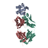
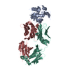
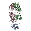
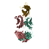
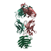
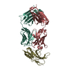
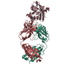
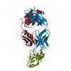
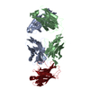
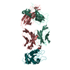
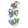
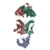
| Entry | Database: PDB / ID: 4tsb | ||||||
|---|---|---|---|---|---|---|---|
| Title | Structure of a lysozyme antibody complex | ||||||
 Components Components |
| ||||||
 Keywords Keywords |  Immune system/hydrolase / antibody / affinity maturation / immune system-hydrolase complex Immune system/hydrolase / antibody / affinity maturation / immune system-hydrolase complex | ||||||
| Function / homology |  Function and homology information Function and homology informationLactose synthesis / Antimicrobial peptides / Neutrophil degranulation / beta-N-acetylglucosaminidase activity / cell wall macromolecule catabolic process / lysozyme / lysozyme activity / killing of cells of another organism / defense response to Gram-negative bacterium / defense response to Gram-positive bacterium ...Lactose synthesis / Antimicrobial peptides / Neutrophil degranulation / beta-N-acetylglucosaminidase activity / cell wall macromolecule catabolic process / lysozyme / lysozyme activity / killing of cells of another organism / defense response to Gram-negative bacterium / defense response to Gram-positive bacterium / defense response to bacterium / endoplasmic reticulum / extracellular space / identical protein binding / cytoplasmSimilarity search - Function | ||||||
| Biological species |  Homo sapiens (human) Homo sapiens (human) Gallus gallus (chicken) Gallus gallus (chicken) | ||||||
| Method | X-RAY DIFFRACTION / SYNCHROTRON / MOLECULAR REPLACEMENT / Resolution: 1.95 Å | ||||||
 Authors Authors | Wensley, B. | ||||||
 Citation Citation | Journal: To Be Published Title: Structure of a lysozyme antibody complex Authors: Wensley, B. | ||||||
| History |
|
- Structure visualization
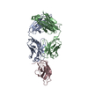
| Structure viewer | Molecule: MolmilJmol/JSmol |
|---|
- Downloads & links
-Download
| PDBx/mmCIF format | 4tsb.cif.gz | 122.9 KB | Display | PDBx/mmCIF format |
|---|---|---|---|---|
| PDB format | pdb4tsb.ent.gz | 97.7 KB | Display | PDB format |
| PDBx/mmJSON format | 4tsb.json.gz | Tree view | PDBx/mmJSON format | |
| Others |  Other downloads Other downloads |
-Validation report
| Arichive directory | https://data.pdbj.org/pub/pdb/validation_reports/ts/4tsbftp://data.pdbj.org/pub/pdb/validation_reports/ts/4tsb | HTTPS FTP |
|---|
-Related structure data


-Links
 PDBj
PDBj


- Assembly
| Deposited unit | 
| ||||||||
|---|---|---|---|---|---|---|---|---|---|
| 1 |
| ||||||||
| Unit cell |
| ||||||||
| Components on special symmetry positions |
|
-Components
| #1: Protein | Mass: 14331.160 Da / Num. of mol.: 1 / Source method: isolated from a natural source / Source: (natural) Gallus gallus (chicken) / References: UniProt: P00698, lysozyme |
|---|---|
| #2: Antibody | Fragment antigen-binding Mass: 23493.297 Da / Num. of mol.: 1 Source method: isolated from a genetically manipulated source Source: (gene. exp.) Homo sapiens (human) / Production host: Homo sapiens (human) |
| #3: Antibody | Fragment antigen-binding Mass: 22826.322 Da / Num. of mol.: 1 Source method: isolated from a genetically manipulated source Source: (gene. exp.) Homo sapiens (human) / Production host: Homo sapiens (human) |
| #4: Water | ChemComp-HOH / Water Mass: 18.015 Da / Num. of mol.: 372 / Source method: isolated from a natural source / Formula: H2O Mass: 18.015 Da / Num. of mol.: 372 / Source method: isolated from a natural source / Formula: H2O |
-Experimental details
-Experiment
| Experiment | Method: X-RAY DIFFRACTION |
|---|
- Sample preparation
| Crystal | Density Matthews: 2.34 Å3/Da / Density % sol: 47.44 % |
|---|---|
| Crystal grow | Temperature: 293 K / Method: vapor diffusion, sitting drop / Details: 100mM PCPT pH 5.5, 200mM MgCl2, 22 % w/v PEG 3350 |
-Data collection
| Diffraction | Mean temperature: 100 K |
|---|---|
| Diffraction source | Source: SYNCHROTRON / Site: Diamond  / Beamline: I04 / Wavelength: 0.979 Å / Beamline: I04 / Wavelength: 0.979 Å |
| Detector | Type: PSI PILATUS 6M / Detector: PIXEL / Date: Mar 11, 2014 |
| Radiation | Protocol: SINGLE WAVELENGTH / Monochromatic (M) / Laue (L): M / Scattering type: x-ray |
| Radiation wavelength | Wavelength: 0.979 Å / Relative weight: 1 |
| Reflection | Resolution: 1.95→34.89 Å / Num. obs: 34734 / % possible obs: 85.4 % / Redundancy: 4.1 % / Biso Wilson estimate: 25.51 Å2 / Net I/σ(I): 12.2 |
| Reflection shell | Resolution: 1.95→2 Å / Redundancy: 2.1 % / Rmerge(I) obs: 0.391 / Mean I/σ(I) obs: 2.2 / % possible all: 39.1 |
- Processing
| Software | Name: BUSTER / Version: 2.11.5 / Classification: refinement | ||||||||||||||||||||||||||||||||||||||||||||||||||||||||||||||||||||||||||||||||||||||||||||||||||||||||||||||||||
|---|---|---|---|---|---|---|---|---|---|---|---|---|---|---|---|---|---|---|---|---|---|---|---|---|---|---|---|---|---|---|---|---|---|---|---|---|---|---|---|---|---|---|---|---|---|---|---|---|---|---|---|---|---|---|---|---|---|---|---|---|---|---|---|---|---|---|---|---|---|---|---|---|---|---|---|---|---|---|---|---|---|---|---|---|---|---|---|---|---|---|---|---|---|---|---|---|---|---|---|---|---|---|---|---|---|---|---|---|---|---|---|---|---|---|---|
| Refinement | Method to determine structure: MOLECULAR REPLACEMENT / Resolution: 1.95→34.89 Å / Cor.coef. Fo:Fc: 0.9344 / Cor.coef. Fo:Fc free: 0.915 / SU R Cruickshank DPI: 0.209 / Cross valid method: THROUGHOUT / σ(F): 0 / SU R Blow DPI: 0.215 / SU Rfree Blow DPI: 0.169 / SU Rfree Cruickshank DPI: 0.168
| ||||||||||||||||||||||||||||||||||||||||||||||||||||||||||||||||||||||||||||||||||||||||||||||||||||||||||||||||||
| Displacement parameters | Biso mean: 23.59 Å2
| ||||||||||||||||||||||||||||||||||||||||||||||||||||||||||||||||||||||||||||||||||||||||||||||||||||||||||||||||||
| Refine analyze | Luzzati coordinate error obs: 0.254 Å | ||||||||||||||||||||||||||||||||||||||||||||||||||||||||||||||||||||||||||||||||||||||||||||||||||||||||||||||||||
| Refinement step | Cycle: 1 / Resolution: 1.95→34.89 Å
| ||||||||||||||||||||||||||||||||||||||||||||||||||||||||||||||||||||||||||||||||||||||||||||||||||||||||||||||||||
| Refine LS restraints |
| ||||||||||||||||||||||||||||||||||||||||||||||||||||||||||||||||||||||||||||||||||||||||||||||||||||||||||||||||||
| LS refinement shell | Resolution: 1.95→2.01 Å / Total num. of bins used: 17
|
