 Movie
Movie Controller

[English] 日本語
 Yorodumi
- PDB-4rxv: The crystal structure of the N-terminal fragment of uncharacteriz... -
+ Open data

- Basic information
| Entry | Database: PDB / ID: 4rxv | ||||||
|---|---|---|---|---|---|---|---|
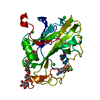
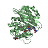
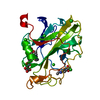
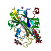
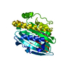
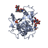
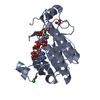
| Title | The crystal structure of the N-terminal fragment of uncharacterized protein from Legionella pneumophila | ||||||
 Components Components | hypothetical protein lpg0944 Hypothesis Hypothesis | ||||||
 Keywords Keywords | STRUCTURAL GENOMICS / UNKNOWN FUNCTION / MCSG / PSI-Biology / Midwest Center for Structural Genomics | ||||||
| Function / homology | Domain of unknown function DUF5617 / Domain of unknown function (DUF5617) / DUF5617 domain-containing protein Function and homology information Function and homology information | ||||||
| Biological species |  Legionella pneumophila subsp. pneumophila str. Philadelphia 1 (bacteria) Legionella pneumophila subsp. pneumophila str. Philadelphia 1 (bacteria) | ||||||
| Method | X-RAY DIFFRACTION / SYNCHROTRON / SAD / Resolution: 1.099 Å | ||||||
 Authors Authors | Nocek, B. / Cuff, M. / Evdokimova, E. / Egorova, O. / Joachimiak, A. / Ensminger, A. / Savchenko, A. / Midwest Center for Structural Genomics (MCSG) | ||||||
 Citation Citation | Journal: Mol Syst Biol / Year: 2016 Title: Diverse mechanisms of metaeffector activity in an intracellular bacterial pathogen, Legionella pneumophila. Authors: Urbanus, M.L. / Quaile, A.T. / Stogios, P.J. / Morar, M. / Rao, C. / Di Leo, R. / Evdokimova, E. / Lam, M. / Oatway, C. / Cuff, M.E. / Osipiuk, J. / Michalska, K. / Nocek, B.P. / Taipale, M. ...Authors: Urbanus, M.L. / Quaile, A.T. / Stogios, P.J. / Morar, M. / Rao, C. / Di Leo, R. / Evdokimova, E. / Lam, M. / Oatway, C. / Cuff, M.E. / Osipiuk, J. / Michalska, K. / Nocek, B.P. / Taipale, M. / Savchenko, A. / Ensminger, A.W. | ||||||
| History |
|
- Structure visualization
| Structure viewer | Molecule: MolmilJmol/JSmol |
|---|
- Downloads & links
-Download
| PDBx/mmCIF format | 4rxv.cif.gz | 154.4 KB | Display | PDBx/mmCIF format |
|---|---|---|---|---|
| PDB format | pdb4rxv.ent.gz | 124.4 KB | Display | PDB format |
| PDBx/mmJSON format | 4rxv.json.gz | Tree view | PDBx/mmJSON format | |
| Others |  Other downloads Other downloads |
-Validation report
| Arichive directory | https://data.pdbj.org/pub/pdb/validation_reports/rx/4rxvftp://data.pdbj.org/pub/pdb/validation_reports/rx/4rxv | HTTPS FTP |
|---|
-Related structure data
| Related structure data | 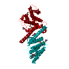 4hfvC 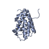 4rxiC 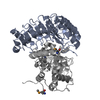 4xa9C  5dggC C: citing same article ( |
|---|---|
| Similar structure data | |
| Other databases |


-Links
 PDBj
PDBj- Assembly
| Deposited unit | 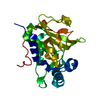
| ||||||||
|---|---|---|---|---|---|---|---|---|---|
| 1 |
| ||||||||
| Unit cell |
|
-Components
| #1: Protein | Hypothesis Mass: 25362.529 Da / Num. of mol.: 1 / Mutation: Q155E Source method: isolated from a genetically manipulated source Source: (gene. exp.) Legionella pneumophila subsp. pneumophila str. Philadelphia 1 (bacteria)Strain: Philadelphia 1 / ATCC 33152 / DSM 7513 / Gene: lpg0944 / Production host: Escherichia coli (E. coli) / References: UniProt: Q5ZWY9 |
|---|---|
| #2: Water | ChemComp-HOH / Water Mass: 18.015 Da / Num. of mol.: 297 / Source method: isolated from a natural source / Formula: H2O Mass: 18.015 Da / Num. of mol.: 297 / Source method: isolated from a natural source / Formula: H2O |
-Experimental details
-Experiment
| Experiment | Method: X-RAY DIFFRACTION / Number of used crystals: 1 |
|---|
- Sample preparation
| Crystal | Density Matthews: 1.72 Å3/Da / Density % sol: 28.6 % |
|---|---|
| Crystal grow | Temperature: 293 K / Method: vapor diffusion, sitting drop / pH: 7.5 Details: 1.6 M ammonium sulfate, 0.1 M NaCl, 0.1 M HEPES pH 7.5, VAPOR DIFFUSION, SITTING DROP, temperature 293K |
-Data collection
| Diffraction | Mean temperature: 100 K |
|---|---|
| Diffraction source | Source: SYNCHROTRON / Site: APS  / Beamline: 19-BM / Wavelength: 0.9794 Å / Beamline: 19-BM / Wavelength: 0.9794 Å |
| Detector | Type: ADSC QUANTUM 315r / Detector: CCD / Date: Feb 13, 2013 / Details: mirrors |
| Radiation | Monochromator: double crystal / Protocol: SINGLE WAVELENGTH / Monochromatic (M) / Laue (L): M / Scattering type: x-ray |
| Radiation wavelength | Wavelength: 0.9794 Å / Relative weight: 1 |
| Reflection | Resolution: 1.099→37.821 Å / Num. all: 69740 / Num. obs: 62764 / % possible obs: 90.1 % / Observed criterion σ(F): 2 / Observed criterion σ(I): 2 / Redundancy: 3.8 % / Rmerge(I) obs: 0.048 / Net I/σ(I): 30 |
| Reflection shell | Resolution: 1.09→1.12 Å / Rmerge(I) obs: 0.05 / % possible all: 45.5 |
- Processing
| Software |
| ||||||||||||||||||||||||||||||||||||||||||||||||||||||||||||||||||||||||||||||||||||||||||||||||||||||||||||||||||||||||||||||||||||||||||||||||||||||||||||||||||||||||
|---|---|---|---|---|---|---|---|---|---|---|---|---|---|---|---|---|---|---|---|---|---|---|---|---|---|---|---|---|---|---|---|---|---|---|---|---|---|---|---|---|---|---|---|---|---|---|---|---|---|---|---|---|---|---|---|---|---|---|---|---|---|---|---|---|---|---|---|---|---|---|---|---|---|---|---|---|---|---|---|---|---|---|---|---|---|---|---|---|---|---|---|---|---|---|---|---|---|---|---|---|---|---|---|---|---|---|---|---|---|---|---|---|---|---|---|---|---|---|---|---|---|---|---|---|---|---|---|---|---|---|---|---|---|---|---|---|---|---|---|---|---|---|---|---|---|---|---|---|---|---|---|---|---|---|---|---|---|---|---|---|---|---|---|---|---|---|---|---|---|
| Refinement | Method to determine structure: SAD / Resolution: 1.099→37.821 Å / SU ML: 0.08 / σ(F): 1.39 / Phase error: 14.45 / Stereochemistry target values: ML
| ||||||||||||||||||||||||||||||||||||||||||||||||||||||||||||||||||||||||||||||||||||||||||||||||||||||||||||||||||||||||||||||||||||||||||||||||||||||||||||||||||||||||
| Solvent computation | Shrinkage radii: 0.9 Å / VDW probe radii: 1.11 Å / Solvent model: FLAT BULK SOLVENT MODEL | ||||||||||||||||||||||||||||||||||||||||||||||||||||||||||||||||||||||||||||||||||||||||||||||||||||||||||||||||||||||||||||||||||||||||||||||||||||||||||||||||||||||||
| Refinement step | Cycle: LAST / Resolution: 1.099→37.821 Å
| ||||||||||||||||||||||||||||||||||||||||||||||||||||||||||||||||||||||||||||||||||||||||||||||||||||||||||||||||||||||||||||||||||||||||||||||||||||||||||||||||||||||||
| Refine LS restraints |
| ||||||||||||||||||||||||||||||||||||||||||||||||||||||||||||||||||||||||||||||||||||||||||||||||||||||||||||||||||||||||||||||||||||||||||||||||||||||||||||||||||||||||
| LS refinement shell |
|
