 Movie
Movie Controller

[English] 日本語
 Yorodumi
- PDB-4rwr: 2.1 Angstrom Crystal Structure of Stage II Sporulation Protein D ... -
+ Open data

- Basic information
| Entry | Database: PDB / ID: 4rwr | ||||||
|---|---|---|---|---|---|---|---|
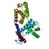
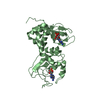
| Title | 2.1 Angstrom Crystal Structure of Stage II Sporulation Protein D from Bacillus anthracis | ||||||
 Components Components | Stage II sporulation protein D | ||||||
 Keywords Keywords |  VIRAL PROTEIN / Structural Genomics / NIAID / National Institute of Allergy and Infectious Diseases / Center for Structural Genomics of Infectious Diseases / CSGID / stage II sporulation protein D / SpoIID / germination VIRAL PROTEIN / Structural Genomics / NIAID / National Institute of Allergy and Infectious Diseases / Center for Structural Genomics of Infectious Diseases / CSGID / stage II sporulation protein D / SpoIID / germination | ||||||
| Function / homology | Sporulation stage II protein D, amidase enhancer LytB / Sporulation stage II protein D, amidase enhancer LytB N-terminal / Sporulation stage II, protein D firmicutes / Stage II sporulation protein / asexual sporulation / sporulation resulting in formation of a cellular spore / Stage II sporulation protein D / Stage II sporulation protein D Function and homology information Function and homology information | ||||||
| Biological species |  Bacillus anthracis (anthrax bacterium) Bacillus anthracis (anthrax bacterium) | ||||||
| Method | X-RAY DIFFRACTION / SYNCHROTRON / SAD / Resolution: 2.1 Å | ||||||
 Authors Authors | Minasov, G. / Wawrzak, Z. / Nocadello, S. / Shuvalova, L. / Dubrovska, I. / Flores, K. / Bagnoli, F. / Falugi, F. / Bottomley, M. / Grandi, G. ...Minasov, G. / Wawrzak, Z. / Nocadello, S. / Shuvalova, L. / Dubrovska, I. / Flores, K. / Bagnoli, F. / Falugi, F. / Bottomley, M. / Grandi, G. / Anderson, W.F. / Center for Structural Genomics of Infectious Diseases (CSGID) | ||||||
 Citation Citation | Journal: J.Biol.Chem. / Year: 2016 Title: Crystal Structures of the SpoIID Lytic Transglycosylases Essential for Bacterial Sporulation. Authors: Nocadello, S. / Minasov, G. / Shuvalova, L.S. / Dubrovska, I. / Sabini, E. / Anderson, W.F. | ||||||
| History |
|
- Structure visualization
| Structure viewer | Molecule: MolmilJmol/JSmol |
|---|
- Downloads & links
-Download
| PDBx/mmCIF format | 4rwr.cif.gz | 230.1 KB | Display | PDBx/mmCIF format |
|---|---|---|---|---|
| PDB format | pdb4rwr.ent.gz | 194 KB | Display | PDB format |
| PDBx/mmJSON format | 4rwr.json.gz | Tree view | PDBx/mmJSON format | |
| Others |  Other downloads Other downloads |
-Validation report
| Arichive directory | https://data.pdbj.org/pub/pdb/validation_reports/rw/4rwrftp://data.pdbj.org/pub/pdb/validation_reports/rw/4rwr | HTTPS FTP |
|---|
-Related structure data


-Links
 PDBj
PDBj- Assembly
| Deposited unit | 
| ||||||||
|---|---|---|---|---|---|---|---|---|---|
| 1 | 
| ||||||||
| 2 | 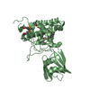
| ||||||||
| Unit cell |
|
-Components
| #1: Protein | Mass: 37055.449 Da / Num. of mol.: 2 / Fragment: UNP residues 32-339 Source method: isolated from a genetically manipulated source Source: (gene. exp.) Bacillus anthracis (anthrax bacterium)Gene: BAS5136, BA_5528, DH23_5378, DJ42_3419, DJ44_3608, DJ45_519, DJ48_4453, DJ49_4581, GBAA_5528, spoIID Plasmid: pMCSG7 / Production host: Escherichia coli (E. coli) / Strain (production host): BL21 Magic / References: UniProt: Q81K14, UniProt: A0A6L7HNW1*PLUS#2: Water | ChemComp-HOH / | Water Mass: 18.015 Da / Num. of mol.: 290 / Source method: isolated from a natural source / Formula: H2O Mass: 18.015 Da / Num. of mol.: 290 / Source method: isolated from a natural source / Formula: H2O |
|---|
-Experimental details
-Experiment
| Experiment | Method: X-RAY DIFFRACTION / Number of used crystals: 1 |
|---|
- Sample preparation
| Crystal | Density Matthews: 1.93 Å3/Da / Density % sol: 36.37 % |
|---|---|
| Crystal grow | Temperature: 295 K / Method: vapor diffusion, sitting drop / pH: 7 Details: Protein: 6.7 mG/mL, 0.5 M Sodium chloride, 0.01 M Tris-HCL buffer, pH 8.3, 2mM N-Acetylglucosamine; Screen: Classics II (H7), 0.15M DL-Malic acid, pH 7.0, 20% (w/v) PEG 3350, VAPOR ...Details: Protein: 6.7 mG/mL, 0.5 M Sodium chloride, 0.01 M Tris-HCL buffer, pH 8.3, 2mM N-Acetylglucosamine; Screen: Classics II (H7), 0.15M DL-Malic acid, pH 7.0, 20% (w/v) PEG 3350, VAPOR DIFFUSION, SITTING DROP, temperature 295K |
-Data collection
| Diffraction | Mean temperature: 100 K |
|---|---|
| Diffraction source | Source: SYNCHROTRON / Site: APS  / Beamline: 21-ID-G / Wavelength: 0.97856 Å / Beamline: 21-ID-G / Wavelength: 0.97856 Å |
| Detector | Type: MARMOSAIC 300 mm CCD / Detector: CCD / Date: Oct 13, 2014 / Details: Beryllium lenses |
| Radiation | Monochromator: Diamond / Protocol: SINGLE WAVELENGTH / Monochromatic (M) / Laue (L): M / Scattering type: x-ray |
| Radiation wavelength | Wavelength: 0.97856 Å / Relative weight: 1 |
| Reflection | Resolution: 2.1→30 Å / Num. all: 32915 / Num. obs: 32915 / % possible obs: 100 % / Observed criterion σ(I): -3 / Redundancy: 4.3 % / Biso Wilson estimate: 29.7 Å2 / Rmerge(I) obs: 0.105 / Rsym value: 0.105 / Net I/σ(I): 12.4 |
| Reflection shell | Resolution: 2.1→2.14 Å / Redundancy: 4.3 % / Rmerge(I) obs: 0.623 / Mean I/σ(I) obs: 2.2 / Num. unique all: 1648 / Rsym value: 0.623 / % possible all: 100 |
- Processing
| Software |
| |||||||||||||||||||||||||||||||||||||||||||||||||||||||||||||||||||||||||||||||||||||||||||||||||||||||||||||||||||||||||||||||||||||||||||||||||||||||||||||||||||||||||||||||||||||||||||||||||||||||||||||||||||||||||||||||||||||||||||||||||||||||||||||||||||||||||||||||||||
|---|---|---|---|---|---|---|---|---|---|---|---|---|---|---|---|---|---|---|---|---|---|---|---|---|---|---|---|---|---|---|---|---|---|---|---|---|---|---|---|---|---|---|---|---|---|---|---|---|---|---|---|---|---|---|---|---|---|---|---|---|---|---|---|---|---|---|---|---|---|---|---|---|---|---|---|---|---|---|---|---|---|---|---|---|---|---|---|---|---|---|---|---|---|---|---|---|---|---|---|---|---|---|---|---|---|---|---|---|---|---|---|---|---|---|---|---|---|---|---|---|---|---|---|---|---|---|---|---|---|---|---|---|---|---|---|---|---|---|---|---|---|---|---|---|---|---|---|---|---|---|---|---|---|---|---|---|---|---|---|---|---|---|---|---|---|---|---|---|---|---|---|---|---|---|---|---|---|---|---|---|---|---|---|---|---|---|---|---|---|---|---|---|---|---|---|---|---|---|---|---|---|---|---|---|---|---|---|---|---|---|---|---|---|---|---|---|---|---|---|---|---|---|---|---|---|---|---|---|---|---|---|---|---|---|---|---|---|---|---|---|---|---|---|---|---|---|---|---|---|---|---|---|---|---|---|---|---|---|---|---|---|---|---|---|---|---|---|---|---|---|---|---|---|---|---|---|
| Refinement | Method to determine structure: SAD / Resolution: 2.1→27.53 Å / Cor.coef. Fo:Fc: 0.953 / Cor.coef. Fo:Fc free: 0.92 / SU B: 10.827 / SU ML: 0.149 Isotropic thermal model: Thermal Factors Individually Isotropically Refined Cross valid method: THROUGHOUT / ESU R: 0.244 / ESU R Free: 0.201 / Stereochemistry target values: MAXIMUM LIKELIHOOD / Details: HYDROGENS HAVE BEEN ADDED IN THE RIDING POSITIONS
| |||||||||||||||||||||||||||||||||||||||||||||||||||||||||||||||||||||||||||||||||||||||||||||||||||||||||||||||||||||||||||||||||||||||||||||||||||||||||||||||||||||||||||||||||||||||||||||||||||||||||||||||||||||||||||||||||||||||||||||||||||||||||||||||||||||||||||||||||||
| Solvent computation | Ion probe radii: 0.8 Å / Shrinkage radii: 0.8 Å / VDW probe radii: 1.2 Å / Solvent model: MASK | |||||||||||||||||||||||||||||||||||||||||||||||||||||||||||||||||||||||||||||||||||||||||||||||||||||||||||||||||||||||||||||||||||||||||||||||||||||||||||||||||||||||||||||||||||||||||||||||||||||||||||||||||||||||||||||||||||||||||||||||||||||||||||||||||||||||||||||||||||
| Displacement parameters | Biso mean: 39.359 Å2
| |||||||||||||||||||||||||||||||||||||||||||||||||||||||||||||||||||||||||||||||||||||||||||||||||||||||||||||||||||||||||||||||||||||||||||||||||||||||||||||||||||||||||||||||||||||||||||||||||||||||||||||||||||||||||||||||||||||||||||||||||||||||||||||||||||||||||||||||||||
| Refinement step | Cycle: LAST / Resolution: 2.1→27.53 Å
| |||||||||||||||||||||||||||||||||||||||||||||||||||||||||||||||||||||||||||||||||||||||||||||||||||||||||||||||||||||||||||||||||||||||||||||||||||||||||||||||||||||||||||||||||||||||||||||||||||||||||||||||||||||||||||||||||||||||||||||||||||||||||||||||||||||||||||||||||||
| Refine LS restraints |
| |||||||||||||||||||||||||||||||||||||||||||||||||||||||||||||||||||||||||||||||||||||||||||||||||||||||||||||||||||||||||||||||||||||||||||||||||||||||||||||||||||||||||||||||||||||||||||||||||||||||||||||||||||||||||||||||||||||||||||||||||||||||||||||||||||||||||||||||||||
| LS refinement shell | Resolution: 2.1→2.154 Å / Total num. of bins used: 20
| |||||||||||||||||||||||||||||||||||||||||||||||||||||||||||||||||||||||||||||||||||||||||||||||||||||||||||||||||||||||||||||||||||||||||||||||||||||||||||||||||||||||||||||||||||||||||||||||||||||||||||||||||||||||||||||||||||||||||||||||||||||||||||||||||||||||||||||||||||
| Refinement TLS params. | Method: refined / Refine-ID: X-RAY DIFFRACTION
| |||||||||||||||||||||||||||||||||||||||||||||||||||||||||||||||||||||||||||||||||||||||||||||||||||||||||||||||||||||||||||||||||||||||||||||||||||||||||||||||||||||||||||||||||||||||||||||||||||||||||||||||||||||||||||||||||||||||||||||||||||||||||||||||||||||||||||||||||||
| Refinement TLS group |
|
