 Movie
Movie Controller


+ Open data

- Basic information
| Entry | Database: PDB / ID: 4rwh | ||||||
|---|---|---|---|---|---|---|---|
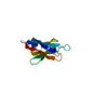
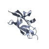
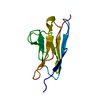
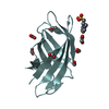
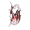
| Title | Crystal structure of T cell costimulatory ligand B7-1 (CD80) | ||||||
 Components Components | T-lymphocyte activation antigen CD80 | ||||||
 Keywords Keywords |  SIGNALING PROTEIN / Ig fold / T cell costimulatory ligand B7-1 SIGNALING PROTEIN / Ig fold / T cell costimulatory ligand B7-1 | ||||||
| Function / homology |  Function and homology information Function and homology informationCD28 dependent PI3K/Akt signaling / CD28 dependent Vav1 pathway / CD28 co-stimulation / CTLA4 inhibitory signaling / protein complex involved in cell adhesion / PIP3 activates AKT signaling / positive regulation of alpha-beta T cell proliferation / PI5P, PP2A and IER3 Regulate PI3K/AKT Signaling / coreceptor activity / positive regulation of T cell proliferation ...CD28 dependent PI3K/Akt signaling / CD28 dependent Vav1 pathway / CD28 co-stimulation / CTLA4 inhibitory signaling / protein complex involved in cell adhesion / PIP3 activates AKT signaling / positive regulation of alpha-beta T cell proliferation / PI5P, PP2A and IER3 Regulate PI3K/AKT Signaling / coreceptor activity / positive regulation of T cell proliferation / negative regulation of T cell proliferation / T cell costimulation / positive regulation of peptidyl-tyrosine phosphorylation / cellular response to lipopolysaccharide / membrane => GO:0016020 / cell surface receptor signaling pathway / immune response / external side of plasma membrane / cell surface / signal transductionSimilarity search - Function | ||||||
| Biological species |  Mus musculus (house mouse) Mus musculus (house mouse) | ||||||
| Method | X-RAY DIFFRACTION / SYNCHROTRON / MOLECULAR REPLACEMENT / Resolution: 1.802 Å | ||||||
 Authors Authors | Fedorov, A.A. / Fedorov, E.V. / Samanta, D. / Hillerich, B. / Seidel, R.D. / Almo, S.C. | ||||||
 Citation Citation | Journal: To be Published Title: Crystal structure of T cell costimulatory ligand B7-1 (CD80) Authors: Fedorov, A.A. / Fedorov, E.V. / Samanta, D. / Hillerich, B. / Seidel, R.D. / Almo, S.C. | ||||||
| History |
|
- Structure visualization
| Structure viewer | Molecule: MolmilJmol/JSmol |
|---|
- Downloads & links
-Download
| PDBx/mmCIF format | 4rwh.cif.gz | 56.6 KB | Display | PDBx/mmCIF format |
|---|---|---|---|---|
| PDB format | pdb4rwh.ent.gz | 40.9 KB | Display | PDB format |
| PDBx/mmJSON format | 4rwh.json.gz | Tree view | PDBx/mmJSON format | |
| Others |  Other downloads Other downloads |
-Validation report
| Arichive directory | https://data.pdbj.org/pub/pdb/validation_reports/rw/4rwhftp://data.pdbj.org/pub/pdb/validation_reports/rw/4rwh | HTTPS FTP |
|---|
-Related structure data
| Related structure data | 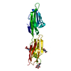 1dr9S S: Starting model for refinement |
|---|---|
| Similar structure data |


-Links
 PDBj
PDBj


- Assembly
| Deposited unit | 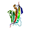
| |||||||||
|---|---|---|---|---|---|---|---|---|---|---|
| 1 |
| |||||||||
| Unit cell |
| |||||||||
| Components on special symmetry positions |
|
-Components
| #1: Protein | Mass: 12335.271 Da / Num. of mol.: 1 / Fragment: UNP residues 44-144 Source method: isolated from a genetically manipulated source Source: (gene. exp.) Mus musculus (house mouse) / Gene: Cd80, B7 / Production host:  Escherichia coli (E. coli) / References: UniProt: Q00609 Escherichia coli (E. coli) / References: UniProt: Q00609 |
|---|---|
| #2: Water | ChemComp-HOH / Water Mass: 18.015 Da / Num. of mol.: 43 / Source method: isolated from a natural source / Formula: H2O Mass: 18.015 Da / Num. of mol.: 43 / Source method: isolated from a natural source / Formula: H2O |
-Experimental details
-Experiment
| Experiment | Method: X-RAY DIFFRACTION / Number of used crystals: 1 |
|---|
- Sample preparation
| Crystal | Density Matthews: 1.81 Å3/Da / Density % sol: 31.88 % |
|---|---|
| Crystal grow | Temperature: 293 K / Method: vapor diffusion, sitting drop / pH: 7 Details: 2.4M sodium malonate, pH 7.0, VAPOR DIFFUSION, SITTING DROP, temperature 293.0K |
-Data collection
| Diffraction | Mean temperature: 100 K |
|---|---|
| Diffraction source | Source: SYNCHROTRON / Site: NSLS  / Beamline: X29A / Wavelength: 1.075 Å / Beamline: X29A / Wavelength: 1.075 Å |
| Detector | Type: ADSC QUANTUM 315r / Detector: CCD / Date: Jul 3, 2014 |
| Radiation | Monochromator: Si 111 CHANNEL / Protocol: SINGLE WAVELENGTH / Monochromatic (M) / Laue (L): M / Scattering type: x-ray |
| Radiation wavelength | Wavelength: 1.075 Å / Relative weight: 1 |
| Reflection | Resolution: 1.802→28.534 Å / Num. all: 11322 / Num. obs: 11322 / % possible obs: 99.46 % / Observed criterion σ(F): 0 / Observed criterion σ(I): 0 |
- Processing
| Software |
| ||||||||||||||||||||||||||||||||||||||||
|---|---|---|---|---|---|---|---|---|---|---|---|---|---|---|---|---|---|---|---|---|---|---|---|---|---|---|---|---|---|---|---|---|---|---|---|---|---|---|---|---|---|
| Refinement | Method to determine structure: MOLECULAR REPLACEMENT Starting model: 1DR9 Resolution: 1.802→28.534 Å / SU ML: 0.15 / σ(F): 0 / Phase error: 31.03 / Stereochemistry target values: ML
| ||||||||||||||||||||||||||||||||||||||||
| Solvent computation | Shrinkage radii: 0.9 Å / VDW probe radii: 1.11 Å / Solvent model: FLAT BULK SOLVENT MODEL | ||||||||||||||||||||||||||||||||||||||||
| Refinement step | Cycle: LAST / Resolution: 1.802→28.534 Å
| ||||||||||||||||||||||||||||||||||||||||
| Refine LS restraints |
| ||||||||||||||||||||||||||||||||||||||||
| LS refinement shell |
| ||||||||||||||||||||||||||||||||||||||||
| Refinement TLS params. | Method: refined / Origin x: 68.0483 Å / Origin y: 11.3089 Å / Origin z: 1.2083 Å
| ||||||||||||||||||||||||||||||||||||||||
| Refinement TLS group | Selection details: chain A |
