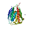
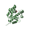
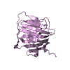
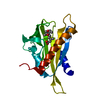
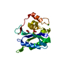
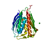
Entry Database : PDB / ID : 4rtiTitle The crystal structure of PsbP from Spinacia oleracea Oxygen-evolving enhancer protein 2, chloroplastic Keywords / / / Function / homology Biological species Spinacia oleracea (spinach)Method / / / Resolution : 1.8 Å Authors Cao, P. / Xie, Y. / Li, M. / Pan, X.W. / Zhang, H.M. / Zhao, X.L. / Su, X.D. / Cheng, T. / Chang, W. Journal : Mol Plant / Year : 2015Title : Crystal structure analysis of extrinsic PsbP protein of photosystem II reveals a manganese-induced conformational change.Authors : Cao, P. / Xie, Y. / Li, M. / Pan, X. / Zhang, H. / Zhao, X. / Su, X. / Cheng, T. / Chang, W. History Deposition Nov 15, 2014 Deposition site / Processing site Revision 1.0 Mar 11, 2015 Provider / Type Revision 1.1 Aug 24, 2022 Group / Derived calculationsCategory citation / citation_author ... citation / citation_author / database_2 / pdbx_struct_conn_angle / struct_conn / struct_site Item _citation.journal_volume / _citation.page_first ... _citation.journal_volume / _citation.page_first / _citation.page_last / _citation.title / _citation_author.name / _database_2.pdbx_DOI / _database_2.pdbx_database_accession / _pdbx_struct_conn_angle.ptnr1_auth_comp_id / _pdbx_struct_conn_angle.ptnr1_auth_seq_id / _pdbx_struct_conn_angle.ptnr1_label_asym_id / _pdbx_struct_conn_angle.ptnr1_label_atom_id / _pdbx_struct_conn_angle.ptnr1_label_comp_id / _pdbx_struct_conn_angle.ptnr1_label_seq_id / _pdbx_struct_conn_angle.ptnr2_auth_seq_id / _pdbx_struct_conn_angle.ptnr2_label_asym_id / _pdbx_struct_conn_angle.ptnr3_auth_comp_id / _pdbx_struct_conn_angle.ptnr3_auth_seq_id / _pdbx_struct_conn_angle.ptnr3_label_asym_id / _pdbx_struct_conn_angle.ptnr3_label_atom_id / _pdbx_struct_conn_angle.ptnr3_label_comp_id / _pdbx_struct_conn_angle.ptnr3_label_seq_id / _pdbx_struct_conn_angle.value / _struct_conn.pdbx_dist_value / _struct_conn.ptnr1_auth_comp_id / _struct_conn.ptnr1_auth_seq_id / _struct_conn.ptnr1_label_asym_id / _struct_conn.ptnr1_label_atom_id / _struct_conn.ptnr1_label_comp_id / _struct_conn.ptnr1_label_seq_id / _struct_conn.ptnr2_auth_comp_id / _struct_conn.ptnr2_auth_seq_id / _struct_conn.ptnr2_label_asym_id / _struct_conn.ptnr2_label_atom_id / _struct_conn.ptnr2_label_comp_id / _struct_site.pdbx_auth_asym_id / _struct_site.pdbx_auth_comp_id / _struct_site.pdbx_auth_seq_id Revision 1.2 Nov 8, 2023 Group / Refinement descriptionCategory / chem_comp_bond / pdbx_initial_refinement_model
Show all Show less
 Movie
Movie Controller


 Open data
 Basic information
 Components
 Keywords
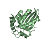
 PHOTOSYNTHESIS / beta-sandwich folding /
  Function and homology information
 Authors
 Citation
 Structure visualization
 Downloads & links
 Other downloads
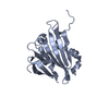


 PDBj
PDBj
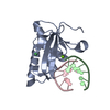
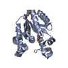
 Assembly
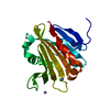

 Mass: 54.938 Da / Num. of mol.: 2 / Source method: obtained synthetically / Formula: Mn
 Mass: 35.453 Da / Num. of mol.: 1 / Source method: obtained synthetically / Formula: Cl
 Mass: 18.015 Da / Num. of mol.: 148 / Source method: isolated from a natural source / Formula: H2O
 Sample preparation
 / Beamline: BL17U / Wavelength: 0.98 Å
 Processing