 Movie
Movie Controller

[English] 日本語
 Yorodumi
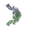
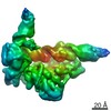
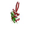
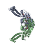
- PDB-4rqf: human Seryl-tRNA synthetase dimer complexed with one molecule of ... -
+ Open data

- Basic information
| Entry | Database: PDB / ID: 4rqf | ||||||
|---|---|---|---|---|---|---|---|
| Title | human Seryl-tRNA synthetase dimer complexed with one molecule of tRNAsec | ||||||
 Components Components |
| ||||||
 Keywords Keywords | LIGASE/RNA /  aminoacyl-tRNA synthetase / classII aaRS / aminoacylation / serine / cytosol / LIGASE-RNA complex aminoacyl-tRNA synthetase / classII aaRS / aminoacylation / serine / cytosol / LIGASE-RNA complex | ||||||
| Function / homology |  Function and homology information Function and homology informationselenocysteine-tRNA ligase activity / negative regulation of vascular endothelial growth factor production / selenocysteine incorporation / seryl-tRNA aminoacylation / serine-tRNA ligase / serine-tRNA ligase activity / Cytosolic tRNA aminoacylation / tRNA modification / Selenocysteine synthesis / negative regulation of angiogenesis ...selenocysteine-tRNA ligase activity / negative regulation of vascular endothelial growth factor production / selenocysteine incorporation / seryl-tRNA aminoacylation / serine-tRNA ligase / serine-tRNA ligase activity / Cytosolic tRNA aminoacylation / tRNA modification / Selenocysteine synthesis / negative regulation of angiogenesis / cytoplasmic translation / tRNA binding / molecular adaptor activity / translation / RNA polymerase II cis-regulatory region sequence-specific DNA binding / enzyme binding / negative regulation of transcription by RNA polymerase II / protein homodimerization activity / extracellular exosome / ATP binding / nucleus / cytosol / cytoplasmSimilarity search - Function | ||||||
| Biological species |  Homo sapiens (human) Homo sapiens (human) | ||||||
| Method | X-RAY DIFFRACTION / SYNCHROTRON / MOLECULAR REPLACEMENT / Resolution: 3.503 Å | ||||||
 Authors Authors | Xie, W. / Wang, C. / Guo, Y. / Tian, Q. / Jia, Q. | ||||||
 Citation Citation | Journal: Nucleic Acids Res. / Year: 2015 Title: SerRS-tRNASec complex structures reveal mechanism of the first step in selenocysteine biosynthesis. Authors: Wang, C. / Guo, Y. / Tian, Q. / Jia, Q. / Gao, Y. / Zhang, Q. / Zhou, C. / Xie, W. | ||||||
| History |
|
- Structure visualization
| Structure viewer | Molecule: MolmilJmol/JSmol |
|---|
- Downloads & links
-Download
| PDBx/mmCIF format | 4rqf.cif.gz | 232.1 KB | Display | PDBx/mmCIF format |
|---|---|---|---|---|
| PDB format | pdb4rqf.ent.gz | 179 KB | Display | PDB format |
| PDBx/mmJSON format | 4rqf.json.gz | Tree view | PDBx/mmJSON format | |
| Others |  Other downloads Other downloads |
-Validation report
| Arichive directory | https://data.pdbj.org/pub/pdb/validation_reports/rq/4rqfftp://data.pdbj.org/pub/pdb/validation_reports/rq/4rqf | HTTPS FTP |
|---|
-Related structure data
| Related structure data |  4rqeC  3a3aS  4l87S C: citing same article ( S: Starting model for refinement |
|---|---|
| Similar structure data |


-Links
 PDBj
PDBj


- Assembly
| Deposited unit | 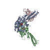
| ||||||||
|---|---|---|---|---|---|---|---|---|---|
| 1 |
| ||||||||
| Unit cell |
|
-Components
| #1: RNA chain | Mass: 28948.107 Da / Num. of mol.: 1 / Mutation: C2G, G70C / Source method: obtained synthetically / Details: in vitro synthesis / Source: (synth.) Homo sapiens (human) | ||||
|---|---|---|---|---|---|
| #2: Protein | Mass: 59934.441 Da / Num. of mol.: 2 / Mutation: E447K Source method: isolated from a genetically manipulated source Source: (gene. exp.) Homo sapiens (human) / Gene: SARS, SERS / Plasmid: pET20b(+) / Production host:  Escherichia coli (E. coli) / Strain (production host): BL21(DE3) / References: UniProt: P49591, serine-tRNA ligase Escherichia coli (E. coli) / Strain (production host): BL21(DE3) / References: UniProt: P49591, serine-tRNA ligase#3: Chemical |   Mass: 506.196 Da / Num. of mol.: 2 / Source method: obtained synthetically / Formula: C10H17N6O12P3 / Comment: AMP-PNP, energy-carrying molecule analogue*YM Mass: 506.196 Da / Num. of mol.: 2 / Source method: obtained synthetically / Formula: C10H17N6O12P3 / Comment: AMP-PNP, energy-carrying molecule analogue*YM#4: Chemical | Serine  Type: L-peptide linking / Mass: 105.093 Da / Num. of mol.: 2 / Source method: obtained synthetically / Formula: C3H7NO3 Type: L-peptide linking / Mass: 105.093 Da / Num. of mol.: 2 / Source method: obtained synthetically / Formula: C3H7NO3 |
-Experimental details
-Experiment
| Experiment | Method: X-RAY DIFFRACTION / Number of used crystals: 1 |
|---|
- Sample preparation
| Crystal | Density Matthews: 2.63 Å3/Da / Density % sol: 53.15 % |
|---|---|
| Crystal grow | Temperature: 298 K / Method: vapor diffusion / pH: 7 Details: 18%(m/v) PEG3350, 0.1M NaCl, 0.1M Tris-HCl (pH8.0), 0.1M Sodium malonate pH7.0., VAPOR DIFFUSION, temperature 298K |
-Data collection
| Diffraction | Mean temperature: 100 K |
|---|---|
| Diffraction source | Source: SYNCHROTRON / Site: SSRF  / Beamline: BL17U / Wavelength: 0.99 Å / Beamline: BL17U / Wavelength: 0.99 Å |
| Detector | Type: ADSC QUANTUM 315r / Detector: CCD / Date: Mar 21, 2014 |
| Radiation | Protocol: SINGLE WAVELENGTH / Monochromatic (M) / Laue (L): M / Scattering type: x-ray |
| Radiation wavelength | Wavelength: 0.99 Å / Relative weight: 1 |
| Reflection | Resolution: 3.503→50 Å / Num. all: 20009 / Num. obs: 19748 / % possible obs: 98.7 % / Observed criterion σ(I): 3 / Redundancy: 5.6 % / Biso Wilson estimate: 115.87 Å2 / Rmerge(I) obs: 0.15 / Net I/σ(I): 12.4 |
| Reflection shell | Resolution: 3.5→3.68 Å / Redundancy: 6.1 % / Rmerge(I) obs: 0.956 / % possible all: 100 |
- Processing
| Software |
| ||||||||||||||||||||||||||||||||||||||||||||||||||||||||
|---|---|---|---|---|---|---|---|---|---|---|---|---|---|---|---|---|---|---|---|---|---|---|---|---|---|---|---|---|---|---|---|---|---|---|---|---|---|---|---|---|---|---|---|---|---|---|---|---|---|---|---|---|---|---|---|---|---|
| Refinement | Method to determine structure: MOLECULAR REPLACEMENT Starting model: 4L87, 3A3A Resolution: 3.503→37.625 Å / SU ML: 0.47 / σ(F): 1.37 / Phase error: 34.14 / Stereochemistry target values: ML
| ||||||||||||||||||||||||||||||||||||||||||||||||||||||||
| Solvent computation | Shrinkage radii: 0.9 Å / VDW probe radii: 1.11 Å / Solvent model: FLAT BULK SOLVENT MODEL | ||||||||||||||||||||||||||||||||||||||||||||||||||||||||
| Refinement step | Cycle: LAST / Resolution: 3.503→37.625 Å
| ||||||||||||||||||||||||||||||||||||||||||||||||||||||||
| Refine LS restraints |
| ||||||||||||||||||||||||||||||||||||||||||||||||||||||||
| LS refinement shell |
|
