 Movie
Movie Controller

[English] 日本語
 Yorodumi
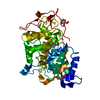
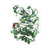
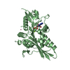
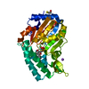
- PDB-4rpf: Crystal structure of homoserine kinase from Yersinia pestis Nepal... -
+ Open data

- Basic information
| Entry | Database: PDB / ID: 4rpf | ||||||
|---|---|---|---|---|---|---|---|
| Title | Crystal structure of homoserine kinase from Yersinia pestis Nepal516, NYSGRC target 032715 | ||||||
 Components Components | Homoserine kinase | ||||||
 Keywords Keywords | TRANSFERASE / PROTEIN STRUCTURE INITIATIVE / PSI-BIOLOGY / NEW YORK STRUCTURAL GENOMICS RESEARCH CONSORTIUM / NYSGRC / KINASE | ||||||
| Function / homology |  Function and homology informationhomoserine kinase / homoserine kinase activity / threonine biosynthetic process / ATP binding / cytoplasm Function and homology informationhomoserine kinase / homoserine kinase activity / threonine biosynthetic process / ATP binding / cytoplasmSimilarity search - Function | ||||||
| Biological species |   Yersinia pestis (bacteria) Yersinia pestis (bacteria) | ||||||
| Method | X-RAY DIFFRACTION / SYNCHROTRON / SAD / Resolution: 2.3 Å | ||||||
 Authors Authors | Ptskovsky, Y. / Bhosle, R. / Toro, R. / Hillerich, B. / Gizzi, A. / Garforth, S. / Kar, A. / Chan, M.K. / Lafluer, J. / Patel, H. ...Ptskovsky, Y. / Bhosle, R. / Toro, R. / Hillerich, B. / Gizzi, A. / Garforth, S. / Kar, A. / Chan, M.K. / Lafluer, J. / Patel, H. / Matikainen, B. / Chamala, S. / Lim, S. / Celikgil, A. / Villegas, G. / Evans, B. / Love, J. / Fiser, A. / Seidel, R. / Bonanno, J.B. / Almo, S.C. / New York Structural Genomics Research Consortium (NYSGRC) | ||||||
 Citation Citation | Journal: To be Published Title: Crystal Structure of Homoserine Kinase from Yersinia Pestis Nepal516, Nysgrc Target 032715 Authors: Patskovsky, Y. / Bhosle, R. / Toro, R. / Hillerich, B. / Gizzi, A. / Garforth, S. / Kar, A. / Chan, M.K. / Lafluer, J. / Patel, H. / Matikainen, B. / Chamala, S. / Lim, S. / Celikgil, A. / ...Authors: Patskovsky, Y. / Bhosle, R. / Toro, R. / Hillerich, B. / Gizzi, A. / Garforth, S. / Kar, A. / Chan, M.K. / Lafluer, J. / Patel, H. / Matikainen, B. / Chamala, S. / Lim, S. / Celikgil, A. / Villegas, G. / Evans, B. / Love, J. / Fiser, A. / Seidel, R. / Bonanno, J.B. / Almo, S.C. / New York Structural Genomics Research Consortium (NYSGRC) | ||||||
| History |
|
- Structure visualization
| Structure viewer | Molecule: MolmilJmol/JSmol |
|---|
- Downloads & links
-Download
| PDBx/mmCIF format | 4rpf.cif.gz | 180.5 KB | Display | PDBx/mmCIF format |
|---|---|---|---|---|
| PDB format | pdb4rpf.ent.gz | 151.5 KB | Display | PDB format |
| PDBx/mmJSON format | 4rpf.json.gz | Tree view | PDBx/mmJSON format | |
| Others |  Other downloads Other downloads |
-Validation report
| Arichive directory | https://data.pdbj.org/pub/pdb/validation_reports/rp/4rpfftp://data.pdbj.org/pub/pdb/validation_reports/rp/4rpf | HTTPS FTP |
|---|
-Related structure data
| Similar structure data | |
|---|---|
| Other databases |


-Links
 PDBj
PDBj- Assembly
| Deposited unit | 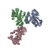
| ||||||||||||||||
|---|---|---|---|---|---|---|---|---|---|---|---|---|---|---|---|---|---|
| 1 | 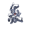
| ||||||||||||||||
| 2 | 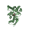
| ||||||||||||||||
| 3 | 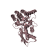
| ||||||||||||||||
| 4 | 
| ||||||||||||||||
| 5 | 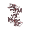
| ||||||||||||||||
| Unit cell |
| ||||||||||||||||
| Noncrystallographic symmetry (NCS) | NCS oper:
|
-Components
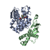
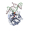
| #1: Protein | / HK / HSK Mass: 33347.250 Da / Num. of mol.: 3 Source method: isolated from a genetically manipulated source Source: (gene. exp.) Yersinia pestis (bacteria) / Strain: Nepal516 / Gene: thrB, YPN_0332, YP516_0338 / Plasmid: PET / Production host: Escherichia coli (E. coli) / Strain (production host): BL21(DE3) / References: UniProt: Q1CMW6, homoserine kinase#2: Chemical | Citric acid  Mass: 192.124 Da / Num. of mol.: 3 / Source method: obtained synthetically / Formula: C6H8O7 Mass: 192.124 Da / Num. of mol.: 3 / Source method: obtained synthetically / Formula: C6H8O7#3: Water | ChemComp-HOH / | Water Mass: 18.015 Da / Num. of mol.: 93 / Source method: isolated from a natural source / Formula: H2O Mass: 18.015 Da / Num. of mol.: 93 / Source method: isolated from a natural source / Formula: H2O |
|---|
-Experimental details
-Experiment
| Experiment | Method: X-RAY DIFFRACTION / Number of used crystals: 1 |
|---|
- Sample preparation
| Crystal | Density Matthews: 4.13 Å3/Da / Density % sol: 70.23 % |
|---|---|
| Crystal grow | Temperature: 294 K / Method: vapor diffusion, sitting drop / pH: 5.6 Details: PROTEIN IN 10 MM BIS-TRIS, 500 MM SODIUM CHLORIDE, 10% GLYCEROL, 5 MM DTT, 10 MM D-GLUCOSE, RESERVOIR: 0.17 M AMMONIUM ACETATE, 0.085 M SODIUM CITRATE, PH 5.6, 25.5% W/V PEG4000, 15% W/V ...Details: PROTEIN IN 10 MM BIS-TRIS, 500 MM SODIUM CHLORIDE, 10% GLYCEROL, 5 MM DTT, 10 MM D-GLUCOSE, RESERVOIR: 0.17 M AMMONIUM ACETATE, 0.085 M SODIUM CITRATE, PH 5.6, 25.5% W/V PEG4000, 15% W/V GLYCEROL, 0.5 M 2-Aminoethylphosphonate; CRYOPROTECTANT = RESERVOIR, VAPOR DIFFUSION, SITTING DROP, TEMPERATURE 294K |
-Data collection
| Diffraction | Mean temperature: 100 K | ||||||||||||||||||||
|---|---|---|---|---|---|---|---|---|---|---|---|---|---|---|---|---|---|---|---|---|---|
| Diffraction source | Source: SYNCHROTRON / Site: NSLS  / Beamline: X29A / Wavelength: 0.979 / Beamline: X29A / Wavelength: 0.979 | ||||||||||||||||||||
| Detector | Type: ADSC QUANTUM 315r / Detector: CCD / Date: Sep 5, 2014 / Details: MIRRORS | ||||||||||||||||||||
| Radiation | Monochromator: SI(111) / Protocol: SINGLE WAVELENGTH / Monochromatic (M) / Laue (L): M / Scattering type: x-ray | ||||||||||||||||||||
| Radiation wavelength | Wavelength: 0.979 Å / Relative weight: 1 | ||||||||||||||||||||
| Reflection twin |
| ||||||||||||||||||||
| Reflection | Resolution: 2.3→50 Å / Num. obs: 70518 / % possible obs: 98.6 % / Observed criterion σ(I): -3 / Redundancy: 6 % / Rmerge(I) obs: 0.084 / Rsym value: 0.084 / Net I/σ(I): 18.25 | ||||||||||||||||||||
| Reflection shell | Resolution: 2.3→2.34 Å / Redundancy: 6 % / Mean I/σ(I) obs: 1.5 / % possible all: 97.9 |
- Processing
| Software |
| ||||||||||||||||||||||||||||||||||||||||||||||||||||||||||||||||||||||||||||||||||||||||||||||||||||||||||||||||||||||||||||||||||||||||||||||||||||||||||||||||||||||||||||||||||||||
|---|---|---|---|---|---|---|---|---|---|---|---|---|---|---|---|---|---|---|---|---|---|---|---|---|---|---|---|---|---|---|---|---|---|---|---|---|---|---|---|---|---|---|---|---|---|---|---|---|---|---|---|---|---|---|---|---|---|---|---|---|---|---|---|---|---|---|---|---|---|---|---|---|---|---|---|---|---|---|---|---|---|---|---|---|---|---|---|---|---|---|---|---|---|---|---|---|---|---|---|---|---|---|---|---|---|---|---|---|---|---|---|---|---|---|---|---|---|---|---|---|---|---|---|---|---|---|---|---|---|---|---|---|---|---|---|---|---|---|---|---|---|---|---|---|---|---|---|---|---|---|---|---|---|---|---|---|---|---|---|---|---|---|---|---|---|---|---|---|---|---|---|---|---|---|---|---|---|---|---|---|---|---|---|
| Refinement | Method to determine structure: SAD Starting model: NONE Resolution: 2.3→40.72 Å / Cor.coef. Fo:Fc: 0.925 / Cor.coef. Fo:Fc free: 0.904 / SU B: 5.054 / SU ML: 0.136 / Cross valid method: THROUGHOUT / ESU R: 0.051 / ESU R Free: 0.047 / Stereochemistry target values: MAXIMUM LIKELIHOOD / Details: HYDROGENS HAVE BEEN ADDED IN THE RIDING POSITIONS
| ||||||||||||||||||||||||||||||||||||||||||||||||||||||||||||||||||||||||||||||||||||||||||||||||||||||||||||||||||||||||||||||||||||||||||||||||||||||||||||||||||||||||||||||||||||||
| Solvent computation | Ion probe radii: 0.8 Å / Shrinkage radii: 0.8 Å / VDW probe radii: 1.1 Å / Solvent model: BABINET MODEL WITH MASK | ||||||||||||||||||||||||||||||||||||||||||||||||||||||||||||||||||||||||||||||||||||||||||||||||||||||||||||||||||||||||||||||||||||||||||||||||||||||||||||||||||||||||||||||||||||||
| Displacement parameters | Biso mean: 57.805 Å2
| ||||||||||||||||||||||||||||||||||||||||||||||||||||||||||||||||||||||||||||||||||||||||||||||||||||||||||||||||||||||||||||||||||||||||||||||||||||||||||||||||||||||||||||||||||||||
| Refinement step | Cycle: LAST / Resolution: 2.3→40.72 Å
| ||||||||||||||||||||||||||||||||||||||||||||||||||||||||||||||||||||||||||||||||||||||||||||||||||||||||||||||||||||||||||||||||||||||||||||||||||||||||||||||||||||||||||||||||||||||
| Refine LS restraints |
|