 Movie
Movie Controller


+ Open data

- Basic information
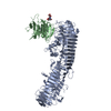
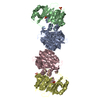
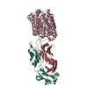
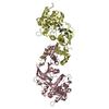
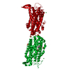
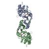
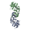
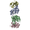
| Entry | Database: PDB / ID: 4rm6 | ||||||
|---|---|---|---|---|---|---|---|
| Title | Crystal structure of Hemopexin Binding Protein | ||||||
 Components Components | Heme/hemopexin-binding protein | ||||||
 Keywords Keywords |  PROTEIN BINDING / beta helix / hemopexin binding protein / hemopexin / Heme-hemopexin-binding protein complex / outer membrane PROTEIN BINDING / beta helix / hemopexin binding protein / hemopexin / Heme-hemopexin-binding protein complex / outer membrane | ||||||
| Function / homology | Filamentous haemagglutinin FhaB/tRNA nuclease CdiA-like, TPS domain / TPS secretion domain / haemagglutination activity domain / Pectin lyase fold / Pectin lyase fold/virulence factor / extracellular region / Heme/hemopexin-binding protein Function and homology information Function and homology information | ||||||
| Biological species |  Haemophilus influenzae Rd KW20 (bacteria) Haemophilus influenzae Rd KW20 (bacteria) | ||||||
| Method | X-RAY DIFFRACTION / SYNCHROTRON / MIR / Resolution: 1.6 Å | ||||||
 Authors Authors | Zambolin, S. / Clantin, B. / Haouz, A. / Villeret, V. / Delepelaire, P. | ||||||
 Citation Citation | Journal: Nat Commun / Year: 2016 Title: Structural basis for haem piracy from host haemopexin by Haemophilus influenzae. Authors: Zambolin, S. / Clantin, B. / Chami, M. / Hoos, S. / Haouz, A. / Villeret, V. / Delepelaire, P. | ||||||
| History |
|
- Structure visualization
| Structure viewer | Molecule: MolmilJmol/JSmol |
|---|
- Downloads & links
-Download
| PDBx/mmCIF format | 4rm6.cif.gz | 321.2 KB | Display | PDBx/mmCIF format |
|---|---|---|---|---|
| PDB format | pdb4rm6.ent.gz | 269.4 KB | Display | PDB format |
| PDBx/mmJSON format | 4rm6.json.gz | Tree view | PDBx/mmJSON format | |
| Others |  Other downloads Other downloads |
-Validation report
| Arichive directory | https://data.pdbj.org/pub/pdb/validation_reports/rm/4rm6ftp://data.pdbj.org/pub/pdb/validation_reports/rm/4rm6 | HTTPS FTP |
|---|
-Related structure data


-Links
 PDBj
PDBj- Assembly
| Deposited unit | 
| ||||||||
|---|---|---|---|---|---|---|---|---|---|
| 1 |
| ||||||||
| Unit cell |
|
-Components
| #1: Protein | Mass: 96594.391 Da / Num. of mol.: 1 / Mutation: C876S, C882S Source method: isolated from a genetically manipulated source Source: (gene. exp.) Haemophilus influenzae Rd KW20 (bacteria)Strain: Rd KW20 / Gene: HI_0264, hxuA / Plasmid: pBAD24 / Production host: Escherichia coli (E. coli) / Strain (production host): BL21 / References: UniProt: P44602 |
|---|---|
| #2: Water | ChemComp-HOH / Water Mass: 18.015 Da / Num. of mol.: 737 / Source method: isolated from a natural source / Formula: H2O Mass: 18.015 Da / Num. of mol.: 737 / Source method: isolated from a natural source / Formula: H2O |
-Experimental details
-Experiment
| Experiment | Method: X-RAY DIFFRACTION / Number of used crystals: 1 |
|---|
- Sample preparation
| Crystal | Density Matthews: 2.35 Å3/Da / Density % sol: 47.71 % |
|---|---|
| Crystal grow | Temperature: 291 K / Method: vapor diffusion, hanging drop / pH: 7.5 Details: Reservoir solution: 0.2 M MgCl2, 0.1 M HEPES pH 7.5, 30% w/v PEG 400, VAPOR DIFFUSION, HANGING DROP, temperature 291K |
-Data collection
| Diffraction | Mean temperature: 100 K | |||||||||||||||||||||||||||||||||||
|---|---|---|---|---|---|---|---|---|---|---|---|---|---|---|---|---|---|---|---|---|---|---|---|---|---|---|---|---|---|---|---|---|---|---|---|---|
| Diffraction source | Source: SYNCHROTRON / Site: SOLEIL  / Beamline: PROXIMA 1 / Wavelength: 0.97918 Å / Beamline: PROXIMA 1 / Wavelength: 0.97918 Å | |||||||||||||||||||||||||||||||||||
| Detector | Type: PSI PILATUS 6M / Detector: PIXEL / Date: Nov 9, 2013 | |||||||||||||||||||||||||||||||||||
| Radiation | Monochromator: cryogenically cooled monochromator crystal / Protocol: SINGLE WAVELENGTH / Monochromatic (M) / Laue (L): M / Scattering type: x-ray | |||||||||||||||||||||||||||||||||||
| Radiation wavelength | Wavelength: 0.97918 Å / Relative weight: 1 | |||||||||||||||||||||||||||||||||||
| Reflection | Resolution: 1.55→30 Å / Num. obs: 131352 / % possible obs: 98.6 % / Observed criterion σ(F): 1 / Observed criterion σ(I): 1 / Biso Wilson estimate: 11.99 Å2 | |||||||||||||||||||||||||||||||||||
| Reflection shell |
|
- Processing
| Software |
| |||||||||||||||||||||||||||||||||||||||||||||||||||||||||||||||||||||||||||
|---|---|---|---|---|---|---|---|---|---|---|---|---|---|---|---|---|---|---|---|---|---|---|---|---|---|---|---|---|---|---|---|---|---|---|---|---|---|---|---|---|---|---|---|---|---|---|---|---|---|---|---|---|---|---|---|---|---|---|---|---|---|---|---|---|---|---|---|---|---|---|---|---|---|---|---|---|
| Refinement | Method to determine structure: MIR / Resolution: 1.6→23.39 Å / Cor.coef. Fo:Fc: 0.8644 / Cor.coef. Fo:Fc free: 0.8581 / SU R Cruickshank DPI: 0.095 / Cross valid method: THROUGHOUT / σ(F): 0 / Stereochemistry target values: Engh & Huber
| |||||||||||||||||||||||||||||||||||||||||||||||||||||||||||||||||||||||||||
| Displacement parameters | Biso mean: 35.56 Å2
| |||||||||||||||||||||||||||||||||||||||||||||||||||||||||||||||||||||||||||
| Refine analyze | Luzzati coordinate error obs: 0.196 Å | |||||||||||||||||||||||||||||||||||||||||||||||||||||||||||||||||||||||||||
| Refinement step | Cycle: LAST / Resolution: 1.6→23.39 Å
| |||||||||||||||||||||||||||||||||||||||||||||||||||||||||||||||||||||||||||
| Refine LS restraints |
| |||||||||||||||||||||||||||||||||||||||||||||||||||||||||||||||||||||||||||
| LS refinement shell | Resolution: 1.6→1.64 Å / Total num. of bins used: 20
| |||||||||||||||||||||||||||||||||||||||||||||||||||||||||||||||||||||||||||
| Refinement TLS params. | Method: refined / Refine-ID: X-RAY DIFFRACTION
| |||||||||||||||||||||||||||||||||||||||||||||||||||||||||||||||||||||||||||
| Refinement TLS group |
|
