 Movie
Movie Controller

[English] 日本語
 Yorodumi
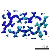
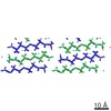
- PDB-4rik: Amyloid forming segment, AVVTGVTAV, from the NAC domain of Parkin... -
+ Open data

- Basic information
| Entry | Database: PDB / ID: 4rik | ||||||
|---|---|---|---|---|---|---|---|
| Title | Amyloid forming segment, AVVTGVTAV, from the NAC domain of Parkinson's disease protein alpha-synuclein, residues 69-77 | ||||||
 Components Components | Alpha-synuclein | ||||||
 Keywords Keywords | LIPID BINDING PROTEIN / Amyloid / alpha-synuclein / Parkinson's Disease / Toxic Core / NAC | ||||||
| Function / homology |  Function and homology information Function and homology informationnegative regulation of mitochondrial electron transport, NADH to ubiquinone / neutral lipid metabolic process / regulation of phospholipase activity / negative regulation of monooxygenase activity / regulation of acyl-CoA biosynthetic process / negative regulation of dopamine uptake involved in synaptic transmission / negative regulation of norepinephrine uptake / positive regulation of glutathione peroxidase activity / positive regulation of SNARE complex assembly / positive regulation of hydrogen peroxide catabolic process ...negative regulation of mitochondrial electron transport, NADH to ubiquinone / neutral lipid metabolic process / regulation of phospholipase activity / negative regulation of monooxygenase activity / regulation of acyl-CoA biosynthetic process / negative regulation of dopamine uptake involved in synaptic transmission / negative regulation of norepinephrine uptake / positive regulation of glutathione peroxidase activity / positive regulation of SNARE complex assembly / positive regulation of hydrogen peroxide catabolic process / supramolecular fiber / negative regulation of transporter activity / negative regulation of chaperone-mediated autophagy / mitochondrial membrane organization / regulation of reactive oxygen species biosynthetic process / positive regulation of protein localization to cell periphery / regulation of synaptic vesicle recycling / negative regulation of platelet-derived growth factor receptor signaling pathway / negative regulation of exocytosis / regulation of glutamate secretion / response to iron(II) ion / regulation of norepinephrine uptake / dopamine biosynthetic process / SNARE complex assembly / positive regulation of neurotransmitter secretion / synaptic vesicle priming / dopamine uptake involved in synaptic transmission / regulation of macrophage activation / regulation of locomotion / positive regulation of inositol phosphate biosynthetic process / mitochondrial ATP synthesis coupled electron transport / negative regulation of microtubule polymerization / synaptic vesicle transport / dynein complex binding / positive regulation of receptor recycling / regulation of dopamine secretion / positive regulation of endocytosis / protein kinase inhibitor activity / negative regulation of thrombin-activated receptor signaling pathway / response to type II interferon / cuprous ion binding / positive regulation of exocytosis / synaptic vesicle exocytosis / cysteine-type endopeptidase inhibitor activity involved in apoptotic process / response to magnesium ion / kinesin binding / alpha-tubulin binding / regulation of presynapse assembly / synaptic vesicle endocytosis / negative regulation of serotonin uptake / localization / phospholipid metabolic process / supramolecular fiber organization / axon terminus / inclusion body / cellular response to copper ion / Hsp70 protein binding / cellular response to epinephrine stimulus / excitatory postsynaptic potential / response to interleukin-1 / adult locomotory behavior / SNARE binding / positive regulation of release of sequestered calcium ion into cytosol / fatty acid metabolic process / long-term synaptic potentiation / regulation of transmembrane transporter activity / ferrous iron binding / synapse organization / phospholipid binding / protein tetramerization / phosphoprotein binding / microglial cell activation / regulation of long-term neuronal synaptic plasticity / negative regulation of protein kinase activity / tau protein binding / protein destabilization / PKR-mediated signaling / negative regulation of cysteine-type endopeptidase activity involved in apoptotic process / positive regulation of protein serine/threonine kinase activity / receptor internalization / synaptic vesicle membrane / positive regulation of inflammatory response / activation of cysteine-type endopeptidase activity involved in apoptotic process / actin cytoskeleton / positive regulation of peptidyl-serine phosphorylation / cellular response to oxidative stress / actin binding / cell cortex / histone binding / growth cone / chemical synaptic transmission / postsynapse / neuron apoptotic process / negative regulation of neuron apoptotic process / amyloid fibril formation / response to lipopolysaccharide / lysosome / molecular adaptor activity / oxidoreductase activity / transcription cis-regulatory region bindingSimilarity search - Function | ||||||
| Biological species |  Homo sapiens (human) Homo sapiens (human) | ||||||
| Method | X-RAY DIFFRACTION / SYNCHROTRON / MOLECULAR REPLACEMENT / molecular replacement / Resolution: 1.854 Å | ||||||
 Authors Authors | Guenther, E.L. / Sawaya, M.R. / Ivanova, M. / Eisenberg, D.S. | ||||||
 Citation Citation | Journal: Nature / Year: 2015 Title: Structure of the toxic core of α-synuclein from invisible crystals. Authors: Jose A Rodriguez / Magdalena I Ivanova / Michael R Sawaya / Duilio Cascio / Francis E Reyes / Dan Shi / Smriti Sangwan / Elizabeth L Guenther / Lisa M Johnson / Meng Zhang / Lin Jiang / Mark ...Authors: Jose A Rodriguez / Magdalena I Ivanova / Michael R Sawaya / Duilio Cascio / Francis E Reyes / Dan Shi / Smriti Sangwan / Elizabeth L Guenther / Lisa M Johnson / Meng Zhang / Lin Jiang / Mark A Arbing / Brent L Nannenga / Johan Hattne / Julian Whitelegge / Aaron S Brewster / Marc Messerschmidt / Sébastien Boutet / Nicholas K Sauter / Tamir Gonen / David S Eisenberg /  Abstract: The protein α-synuclein is the main component of Lewy bodies, the neuron-associated aggregates seen in Parkinson disease and other neurodegenerative pathologies. An 11-residue segment, which we term ...The protein α-synuclein is the main component of Lewy bodies, the neuron-associated aggregates seen in Parkinson disease and other neurodegenerative pathologies. An 11-residue segment, which we term NACore, appears to be responsible for amyloid formation and cytotoxicity of human α-synuclein. Here we describe crystals of NACore that have dimensions smaller than the wavelength of visible light and thus are invisible by optical microscopy. As the crystals are thousands of times too small for structure determination by synchrotron X-ray diffraction, we use micro-electron diffraction to determine the structure at atomic resolution. The 1.4 Å resolution structure demonstrates that this method can determine previously unknown protein structures and here yields, to our knowledge, the highest resolution achieved by any cryo-electron microscopy method to date. The structure exhibits protofibrils built of pairs of face-to-face β-sheets. X-ray fibre diffraction patterns show the similarity of NACore to toxic fibrils of full-length α-synuclein. The NACore structure, together with that of a second segment, inspires a model for most of the ordered portion of the toxic, full-length α-synuclein fibril, presenting opportunities for the design of inhibitors of α-synuclein fibrils. | ||||||
| History |
|
- Structure visualization
| Structure viewer | Molecule: MolmilJmol/JSmol |
|---|
- Downloads & links
-Download
| PDBx/mmCIF format | 4rik.cif.gz | 9 KB | Display | PDBx/mmCIF format |
|---|---|---|---|---|
| PDB format | pdb4rik.ent.gz | 5.5 KB | Display | PDB format |
| PDBx/mmJSON format | 4rik.json.gz | Tree view | PDBx/mmJSON format | |
| Others |  Other downloads Other downloads |
-Validation report
| Arichive directory | https://data.pdbj.org/pub/pdb/validation_reports/ri/4rikftp://data.pdbj.org/pub/pdb/validation_reports/ri/4rik | HTTPS FTP |
|---|
-Related structure data


-Links
 PDBj
PDBj
- Assembly
| Deposited unit | 
| ||||||||
|---|---|---|---|---|---|---|---|---|---|
| 1 | x 10
| ||||||||
| Unit cell |
| ||||||||
| Details | The biological unit is a pair of beta-sheets. One sheet is composed of chain A and unit cell translations along the b dimension (for example, x,y+1,z, etc.). The other sheet is composed of the symmetry mate -x+1/2,y+1/2,-z+1, and its unit cell translations along b (for example, -x+1/2,y+3/2,-z+1, etc.). |
-Components
| #1: Protein/peptide | / Non-A beta component of AD amyloid / Non-A4 component of amyloid precursor / NACP Mass: 815.953 Da / Num. of mol.: 1 / Source method: obtained synthetically Details: Synthetic peptide AVVTGVTAV corresponding to segment 69-77 of human alpha-synuclein Source: (synth.) Homo sapiens (human) / References: UniProt: P37840 |
|---|---|
| #2: Water | ChemComp-HOH / Water Mass: 18.015 Da / Num. of mol.: 3 / Source method: isolated from a natural source / Formula: H2O Mass: 18.015 Da / Num. of mol.: 3 / Source method: isolated from a natural source / Formula: H2O |
-Experimental details
-Experiment
| Experiment | Method: X-RAY DIFFRACTION / Number of used crystals: 1 |
|---|
- Sample preparation
| Crystal | Density Matthews: 1.52 Å3/Da / Density % sol: 19.22 % |
|---|---|
| Crystal grow | Temperature: 273 K / Method: vapor diffusion, hanging drop / pH: 4.6 Details: 0.9M Ammonium Phosphate, 0.1M Sodium Acetate, pH 4.6, vapor diffusion, hanging drop, temperature 273K |
-Data collection
| Diffraction | Mean temperature: 100 K | |||||||||||||||||||||||||||||||||||||||||||||||||||||||||||||||||||||||||||||
|---|---|---|---|---|---|---|---|---|---|---|---|---|---|---|---|---|---|---|---|---|---|---|---|---|---|---|---|---|---|---|---|---|---|---|---|---|---|---|---|---|---|---|---|---|---|---|---|---|---|---|---|---|---|---|---|---|---|---|---|---|---|---|---|---|---|---|---|---|---|---|---|---|---|---|---|---|---|---|
| Diffraction source | Source: SYNCHROTRON / Site: APS / Beamline: 24-ID-E / Wavelength: 0.9791 Å | |||||||||||||||||||||||||||||||||||||||||||||||||||||||||||||||||||||||||||||
| Detector | Type: ADSC QUANTUM 315 / Detector: CCD / Date: Aug 23, 2011 | |||||||||||||||||||||||||||||||||||||||||||||||||||||||||||||||||||||||||||||
| Radiation | Protocol: SINGLE WAVELENGTH / Monochromatic (M) / Laue (L): M / Scattering type: x-ray | |||||||||||||||||||||||||||||||||||||||||||||||||||||||||||||||||||||||||||||
| Radiation wavelength | Wavelength: 0.9791 Å / Relative weight: 1 | |||||||||||||||||||||||||||||||||||||||||||||||||||||||||||||||||||||||||||||
| Reflection | Resolution: 1.85→100 Å / Num. all: 521 / Num. obs: 521 / % possible obs: 97.7 % / Observed criterion σ(I): -3 / Redundancy: 4.1 % / Rmerge(I) obs: 0.117 / Χ2: 0.961 / Net I/σ(I): 11.1 | |||||||||||||||||||||||||||||||||||||||||||||||||||||||||||||||||||||||||||||
| Reflection shell |
|
-Phasing
| Phasing | Method: molecular replacement |
|---|---|
| Phasing MR | Model details: Phaser MODE: MR_AUTO |
- Processing
| Software |
| |||||||||||||||||||||||||||||||||||||||||||||||||||||||||||||||||
|---|---|---|---|---|---|---|---|---|---|---|---|---|---|---|---|---|---|---|---|---|---|---|---|---|---|---|---|---|---|---|---|---|---|---|---|---|---|---|---|---|---|---|---|---|---|---|---|---|---|---|---|---|---|---|---|---|---|---|---|---|---|---|---|---|---|---|
| Refinement | Method to determine structure: MOLECULAR REPLACEMENT Starting model: poly alanine Resolution: 1.854→30 Å / Cor.coef. Fo:Fc: 0.978 / Cor.coef. Fo:Fc free: 0.961 / WRfactor Rfree: 0.236 / WRfactor Rwork: 0.19 / FOM work R set: 0.73 / SU B: 4.061 / SU ML: 0.112 / SU R Cruickshank DPI: 0.1735 / SU Rfree: 0.1526 / Cross valid method: THROUGHOUT / σ(F): 0 / ESU R: 0.173 / ESU R Free: 0.153 / Stereochemistry target values: MAXIMUM LIKELIHOOD Details: HYDROGENS HAVE BEEN ADDED IN THE RIDING POSITIONS U VALUES : REFINED INDIVIDUALLY
| |||||||||||||||||||||||||||||||||||||||||||||||||||||||||||||||||
| Solvent computation | Ion probe radii: 0.8 Å / Shrinkage radii: 0.8 Å / VDW probe radii: 1.2 Å / Solvent model: MASK | |||||||||||||||||||||||||||||||||||||||||||||||||||||||||||||||||
| Displacement parameters | Biso max: 53.1 Å2 / Biso mean: 17.341 Å2 / Biso min: 6.25 Å2
| |||||||||||||||||||||||||||||||||||||||||||||||||||||||||||||||||
| Refinement step | Cycle: LAST / Resolution: 1.854→30 Å /
| |||||||||||||||||||||||||||||||||||||||||||||||||||||||||||||||||
| Refine LS restraints |
| |||||||||||||||||||||||||||||||||||||||||||||||||||||||||||||||||
| LS refinement shell | Resolution: 1.854→2.072 Å / Total num. of bins used: 5
|
