 Movie
Movie Controller


+ Open data

- Basic information
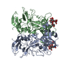
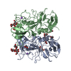
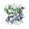
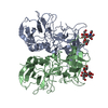
| Entry | Database: PDB / ID: 4rdj | ||||||
|---|---|---|---|---|---|---|---|
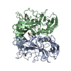
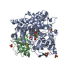
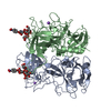
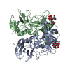
| Title | Crystal structure of Norovirus Boxer P domain | ||||||
 Components Components | Capsid | ||||||
 Keywords Keywords | VIRAL PROTEIN / mixed alpha/beta structure / virus capsid / receptor binding / HBGA | ||||||
| Function / homology |  Function and homology information Function and homology informationPositive stranded ssRNA viruses / Nucleoplasmin-like/VP (viral coat and capsid proteins) / Positive stranded ssRNA viruses / Calicivirus coat protein C-terminal / Calicivirus coat protein C-terminal / Calicivirus coat protein / Calicivirus coat protein / Elongation Factor Tu (Ef-tu); domain 3 / Viral coat protein subunit / Beta Barrel / Mainly BetaSimilarity search - Domain/homology | ||||||
| Biological species |  Human calicivirus NLV/Boxer/2001/US Human calicivirus NLV/Boxer/2001/US | ||||||
| Method | X-RAY DIFFRACTION / SYNCHROTRON / MOLECULAR REPLACEMENT / Resolution: 1.5 Å | ||||||
 Authors Authors | Hao, N. / Chen, Y. / Xia, M. / Liu, W. / Tan, M. / Jiang, X. / Li, X. | ||||||
 Citation Citation | Journal: Protein Cell / Year: 2015 Title: Crystal structures of GI.8 Boxer virus P dimers in complex with HBGAs, a novel evolutionary path selected by the Lewis epitope. Authors: Hao, N. / Chen, Y. / Xia, M. / Tan, M. / Liu, W. / Guan, X. / Jiang, X. / Li, X. / Rao, Z. | ||||||
| History |
|
- Structure visualization
| Structure viewer | Molecule: MolmilJmol/JSmol |
|---|
- Downloads & links
-Download
| PDBx/mmCIF format | 4rdj.cif.gz | 254.3 KB | Display | PDBx/mmCIF format |
|---|---|---|---|---|
| PDB format | pdb4rdj.ent.gz | 204.1 KB | Display | PDB format |
| PDBx/mmJSON format | 4rdj.json.gz | Tree view | PDBx/mmJSON format | |
| Others |  Other downloads Other downloads |
-Validation report
| Arichive directory | https://data.pdbj.org/pub/pdb/validation_reports/rd/4rdjftp://data.pdbj.org/pub/pdb/validation_reports/rd/4rdj | HTTPS FTP |
|---|
-Related structure data
| Related structure data |  4rdkC  4rdlC  2zl7S C: citing same article ( S: Starting model for refinement |
|---|---|
| Similar structure data |


-Links
 PDBj
PDBj
- Assembly
| Deposited unit | 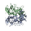
| ||||||||
|---|---|---|---|---|---|---|---|---|---|
| 1 |
| ||||||||
| Unit cell |
|
-Components
| #1: Protein | Mass: 33758.879 Da / Num. of mol.: 2 / Fragment: Protrusion domain, UNP residues 227-526 Source method: isolated from a genetically manipulated source Source: (gene. exp.) Human calicivirus NLV/Boxer/2001/US / Plasmid: pGex-6p-1 / Production host:  Escherichia coli BL21(DE3) (bacteria) / References: UniProt: Q8BCA3 Escherichia coli BL21(DE3) (bacteria) / References: UniProt: Q8BCA3#2: Water | ChemComp-HOH / | Water Mass: 18.015 Da / Num. of mol.: 764 / Source method: isolated from a natural source / Formula: H2O Mass: 18.015 Da / Num. of mol.: 764 / Source method: isolated from a natural source / Formula: H2O |
|---|
-Experimental details
-Experiment
| Experiment | Method: X-RAY DIFFRACTION / Number of used crystals: 1 |
|---|
- Sample preparation
| Crystal | Density Matthews: 2.71 Å3/Da / Density % sol: 54.57 % |
|---|---|
| Crystal grow | Temperature: 290 K / Method: vapor diffusion, hanging drop / pH: 7.5 Details: 0.1M LiCl, 18% (w/v) PEG 3350, 10% (v/v) MPD, pH 7.5, VAPOR DIFFUSION, HANGING DROP, temperature 290K |
-Data collection
| Diffraction | Mean temperature: 100 K |
|---|---|
| Diffraction source | Source: SYNCHROTRON / Site: Photon Factory  / Beamline: BL-17A / Wavelength: 1 Å / Beamline: BL-17A / Wavelength: 1 Å |
| Detector | Type: ADSC QUANTUM 315r / Detector: CCD / Date: May 28, 2012 / Details: mirrors |
| Radiation | Monochromator: Si(111) / Protocol: SINGLE WAVELENGTH / Monochromatic (M) / Laue (L): M / Scattering type: x-ray |
| Radiation wavelength | Wavelength: 1 Å / Relative weight: 1 |
| Reflection | Resolution: 1.5→50 Å / Num. all: 115571 / Num. obs: 115571 / % possible obs: 100 % / Observed criterion σ(F): 2 / Observed criterion σ(I): 2 / Redundancy: 11.2 % / Rmerge(I) obs: 0.047 / Net I/σ(I): 42.9 |
| Reflection shell | Resolution: 1.5→1.53 Å / Redundancy: 11 % / Rmerge(I) obs: 0.419 / Mean I/σ(I) obs: 5.4 / % possible all: 100 |
- Processing
| Software |
| |||||||||||||||||||||||||||||||||||||||||||||||||||||||||||||||||||||||||||||||||||||||||||||||||||||||||||||||||||||||||||||||||||||||||||||||||||||||||||
|---|---|---|---|---|---|---|---|---|---|---|---|---|---|---|---|---|---|---|---|---|---|---|---|---|---|---|---|---|---|---|---|---|---|---|---|---|---|---|---|---|---|---|---|---|---|---|---|---|---|---|---|---|---|---|---|---|---|---|---|---|---|---|---|---|---|---|---|---|---|---|---|---|---|---|---|---|---|---|---|---|---|---|---|---|---|---|---|---|---|---|---|---|---|---|---|---|---|---|---|---|---|---|---|---|---|---|---|---|---|---|---|---|---|---|---|---|---|---|---|---|---|---|---|---|---|---|---|---|---|---|---|---|---|---|---|---|---|---|---|---|---|---|---|---|---|---|---|---|---|---|---|---|---|---|---|---|
| Refinement | Method to determine structure: MOLECULAR REPLACEMENT Starting model: PDB entry 2ZL7 Resolution: 1.5→29.377 Å / SU ML: 0.15 / Isotropic thermal model: anisotropic / Cross valid method: THROUGHOUT / σ(F): 1.34 / Phase error: 14.31 / Stereochemistry target values: ML
| |||||||||||||||||||||||||||||||||||||||||||||||||||||||||||||||||||||||||||||||||||||||||||||||||||||||||||||||||||||||||||||||||||||||||||||||||||||||||||
| Solvent computation | Shrinkage radii: 0.95 Å / VDW probe radii: 1.2 Å / Solvent model: FLAT BULK SOLVENT MODEL / Bsol: 35.44 Å2 / ksol: 0.34 e/Å3 | |||||||||||||||||||||||||||||||||||||||||||||||||||||||||||||||||||||||||||||||||||||||||||||||||||||||||||||||||||||||||||||||||||||||||||||||||||||||||||
| Displacement parameters | Biso mean: 19.9 Å2
| |||||||||||||||||||||||||||||||||||||||||||||||||||||||||||||||||||||||||||||||||||||||||||||||||||||||||||||||||||||||||||||||||||||||||||||||||||||||||||
| Refinement step | Cycle: LAST / Resolution: 1.5→29.377 Å
| |||||||||||||||||||||||||||||||||||||||||||||||||||||||||||||||||||||||||||||||||||||||||||||||||||||||||||||||||||||||||||||||||||||||||||||||||||||||||||
| Refine LS restraints |
| |||||||||||||||||||||||||||||||||||||||||||||||||||||||||||||||||||||||||||||||||||||||||||||||||||||||||||||||||||||||||||||||||||||||||||||||||||||||||||
| LS refinement shell | Refine-ID: X-RAY DIFFRACTION / Total num. of bins used: 30 / % reflection obs: 100 %
|
