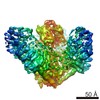
 Movie
Movie Controller


+ Open data

- Basic information
| Entry | Database: PDB / ID: 4rax | ||||||
|---|---|---|---|---|---|---|---|
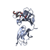
| Title | A regulatory domain of an ion channel | ||||||
 Components Components | Piezo-type mechanosensitive ion channel component 1 | ||||||
 Keywords Keywords |  STRUCTURAL PROTEIN / sandwich fold / novel structure / extra-cellular / regulatory domain / regulatory STRUCTURAL PROTEIN / sandwich fold / novel structure / extra-cellular / regulatory domain / regulatory | ||||||
| Function / homology |  Function and homology information Function and homology informationmechanosensitive monoatomic cation channel activity / positive regulation of cell-cell adhesion mediated by integrin / detection of mechanical stimulus / mechanosensitive monoatomic ion channel activity / positive regulation of integrin activation / positive regulation of myotube differentiation / lamellipodium membrane / monoatomic cation transport / monoatomic cation channel activity / regulation of membrane potential ...mechanosensitive monoatomic cation channel activity / positive regulation of cell-cell adhesion mediated by integrin / detection of mechanical stimulus / mechanosensitive monoatomic ion channel activity / positive regulation of integrin activation / positive regulation of myotube differentiation / lamellipodium membrane / monoatomic cation transport / monoatomic cation channel activity / regulation of membrane potential / endoplasmic reticulum-Golgi intermediate compartment membrane / cellular response to mechanical stimulus / endoplasmic reticulum membrane / endoplasmic reticulum / identical protein binding / plasma membraneSimilarity search - Function | ||||||
| Biological species |  Mus musculus (house mouse) Mus musculus (house mouse) | ||||||
| Method | X-RAY DIFFRACTION / SYNCHROTRON / SAD / Resolution: 1.45 Å | ||||||
 Authors Authors | Ge, J. / Yang, M. | ||||||
 Citation Citation | Journal: Nature / Year: 2015 Title: Architecture of the mammalian mechanosensitive Piezo1 channel. Authors: Jingpeng Ge / Wanqiu Li / Qiancheng Zhao / Ningning Li / Maofei Chen / Peng Zhi / Ruochong Li / Ning Gao / Bailong Xiao / Maojun Yang /  Abstract: Piezo proteins are evolutionarily conserved and functionally diverse mechanosensitive cation channels. However, the overall structural architecture and gating mechanisms of Piezo channels have ...Piezo proteins are evolutionarily conserved and functionally diverse mechanosensitive cation channels. However, the overall structural architecture and gating mechanisms of Piezo channels have remained unknown. Here we determine the cryo-electron microscopy structure of the full-length (2,547 amino acids) mouse Piezo1 (Piezo1) at a resolution of 4.8 Å. Piezo1 forms a trimeric propeller-like structure (about 900 kilodalton), with the extracellular domains resembling three distal blades and a central cap. The transmembrane region has 14 apparently resolved segments per subunit. These segments form three peripheral wings and a central pore module that encloses a potential ion-conducting pore. The rather flexible extracellular blade domains are connected to the central intracellular domain by three long beam-like structures. This trimeric architecture suggests that Piezo1 may use its peripheral regions as force sensors to gate the central ion-conducting pore. | ||||||
| History |
|
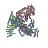
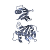
- Structure visualization
| Structure viewer | Molecule: MolmilJmol/JSmol |
|---|
- Downloads & links
-Download
| PDBx/mmCIF format | 4rax.cif.gz | 107.6 KB | Display | PDBx/mmCIF format |
|---|---|---|---|---|
| PDB format | pdb4rax.ent.gz | 86.2 KB | Display | PDB format |
| PDBx/mmJSON format | 4rax.json.gz | Tree view | PDBx/mmJSON format | |
| Others |  Other downloads Other downloads |
-Validation report
| Arichive directory | https://data.pdbj.org/pub/pdb/validation_reports/ra/4raxftp://data.pdbj.org/pub/pdb/validation_reports/ra/4rax | HTTPS FTP |
|---|
-Related structure data


-Links
 PDBj
PDBj
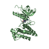
- Assembly
| Deposited unit | 
| ||||||||
|---|---|---|---|---|---|---|---|---|---|
| 1 |
| ||||||||
| Unit cell |
|
-Components
| #1: Protein | Mass: 28346.787 Da / Num. of mol.: 1 / Fragment: UNP residues 2214-2457 Source method: isolated from a genetically manipulated source Source: (gene. exp.) Mus musculus (house mouse) / Gene: Piezo1, Fam38a / Production host:  Escherichia coli (E. coli) / References: UniProt: E2JF22 Escherichia coli (E. coli) / References: UniProt: E2JF22 |
|---|---|
| #2: Water | ChemComp-HOH / Water Mass: 18.015 Da / Num. of mol.: 352 / Source method: isolated from a natural source / Formula: H2O Mass: 18.015 Da / Num. of mol.: 352 / Source method: isolated from a natural source / Formula: H2O |
-Experimental details
-Experiment
| Experiment | Method: X-RAY DIFFRACTION / Number of used crystals: 1 |
|---|
- Sample preparation
| Crystal | Density Matthews: 2.11 Å3/Da / Density % sol: 41.68 % |
|---|---|
| Crystal grow | Temperature: 291 K / Method: vapor diffusion, sitting drop / pH: 7.5 Details: 0.2M Magnesium chloride, 0.1M Hepes pH 7.5, 25% w/w PEG3350, VAPOR DIFFUSION, SITTING DROP, temperature 291K |
-Data collection
| Diffraction | Mean temperature: 100 K | |||||||||||||||||||||||||||||||||
|---|---|---|---|---|---|---|---|---|---|---|---|---|---|---|---|---|---|---|---|---|---|---|---|---|---|---|---|---|---|---|---|---|---|---|
| Diffraction source | Source: SYNCHROTRON / Site: SSRF / Beamline: BL17U / Wavelength: 0.9792 Å | |||||||||||||||||||||||||||||||||
| Detector | Type: ADSC QUANTUM 315 / Detector: CCD / Date: Apr 15, 2014 | |||||||||||||||||||||||||||||||||
| Radiation | Protocol: SINGLE WAVELENGTH / Monochromatic (M) / Laue (L): M / Scattering type: x-ray | |||||||||||||||||||||||||||||||||
| Radiation wavelength | Wavelength: 0.9792 Å / Relative weight: 1 | |||||||||||||||||||||||||||||||||
| Reflection | Resolution: 1.45→50 Å / Num. all: 42523 / Num. obs: 42493 / % possible obs: 99.9 % / Redundancy: 7.6 % / Rmerge(I) obs: 0.079 / Rsym value: 0.107 | |||||||||||||||||||||||||||||||||
| Reflection shell |
|
- Processing
| Software |
| ||||||||||||||||||||||||||||||||||||||||||||||||||||||||||||||||||||||||||||||||||||||||||||||||||||||||||||||||
|---|---|---|---|---|---|---|---|---|---|---|---|---|---|---|---|---|---|---|---|---|---|---|---|---|---|---|---|---|---|---|---|---|---|---|---|---|---|---|---|---|---|---|---|---|---|---|---|---|---|---|---|---|---|---|---|---|---|---|---|---|---|---|---|---|---|---|---|---|---|---|---|---|---|---|---|---|---|---|---|---|---|---|---|---|---|---|---|---|---|---|---|---|---|---|---|---|---|---|---|---|---|---|---|---|---|---|---|---|---|---|---|---|---|
| Refinement | Method to determine structure: SAD / Resolution: 1.45→36.535 Å / SU ML: 0.13 / σ(F): 1.34 / Phase error: 16.64 / Stereochemistry target values: ML
| ||||||||||||||||||||||||||||||||||||||||||||||||||||||||||||||||||||||||||||||||||||||||||||||||||||||||||||||||
| Solvent computation | Shrinkage radii: 0.9 Å / VDW probe radii: 1.11 Å / Solvent model: FLAT BULK SOLVENT MODEL | ||||||||||||||||||||||||||||||||||||||||||||||||||||||||||||||||||||||||||||||||||||||||||||||||||||||||||||||||
| Refinement step | Cycle: LAST / Resolution: 1.45→36.535 Å
| ||||||||||||||||||||||||||||||||||||||||||||||||||||||||||||||||||||||||||||||||||||||||||||||||||||||||||||||||
| Refine LS restraints |
| ||||||||||||||||||||||||||||||||||||||||||||||||||||||||||||||||||||||||||||||||||||||||||||||||||||||||||||||||
| LS refinement shell |
| ||||||||||||||||||||||||||||||||||||||||||||||||||||||||||||||||||||||||||||||||||||||||||||||||||||||||||||||||
| Refinement TLS params. | Method: refined / Origin x: -1.2043 Å / Origin y: 16.0277 Å / Origin z: 34.498 Å
| ||||||||||||||||||||||||||||||||||||||||||||||||||||||||||||||||||||||||||||||||||||||||||||||||||||||||||||||||
| Refinement TLS group | Selection details: all |
