 Movie
Movie Controller

[English] 日本語
 Yorodumi
- PDB-4ra9: Crystal Structure of Conjoint Pyrococcus Furiosus L-asparaginase ... -
+ Open data

- Basic information
| Entry | Database: PDB / ID: 4ra9 | ||||||
|---|---|---|---|---|---|---|---|
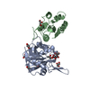
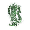
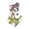
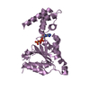
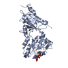
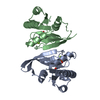
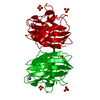
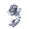
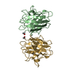
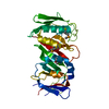
| Title | Crystal Structure of Conjoint Pyrococcus Furiosus L-asparaginase with Citrate | ||||||
 Components Components | (L-asparaginase Asparaginase) x 2 Asparaginase) x 2 | ||||||
 Keywords Keywords | HYDROLASE | ||||||
| Function / homology |  Function and homology informationasparaginase / asparaginase activity / amino acid metabolic process / cytosol Function and homology informationasparaginase / asparaginase activity / amino acid metabolic process / cytosolSimilarity search - Function | ||||||
| Biological species |   Pyrococcus furiosus DSM 3638 (archaea) Pyrococcus furiosus DSM 3638 (archaea) | ||||||
| Method | X-RAY DIFFRACTION / MOLECULAR REPLACEMENT / Resolution: 2.049 Å | ||||||
 Authors Authors | Sharma, P. / Tomar, R. / Singh, S. / Yadav, S.P.S. / Ashish / Kundu, B. | ||||||
 Citation Citation | Journal: Acta Crystallogr.,Sect.D / Year: 2014 Title: Structural and functional insights into an archaeal L-asparaginase obtained through the linker-less assembly of constituent domains. Authors: Tomar, R. / Sharma, P. / Srivastava, A. / Bansal, S. / Kundu, B. | ||||||
| History |
|
- Structure visualization
| Structure viewer | Molecule: MolmilJmol/JSmol |
|---|
- Downloads & links
-Download
| PDBx/mmCIF format | 4ra9.cif.gz | 81.1 KB | Display | PDBx/mmCIF format |
|---|---|---|---|---|
| PDB format | pdb4ra9.ent.gz | 59.4 KB | Display | PDB format |
| PDBx/mmJSON format | 4ra9.json.gz | Tree view | PDBx/mmJSON format | |
| Others |  Other downloads Other downloads |
-Validation report
| Arichive directory | https://data.pdbj.org/pub/pdb/validation_reports/ra/4ra9ftp://data.pdbj.org/pub/pdb/validation_reports/ra/4ra9 | HTTPS FTP |
|---|
-Related structure data
| Related structure data |  4njeSC 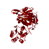 4q0mC 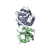 4ra6C C: citing same article ( S: Starting model for refinement |
|---|---|
| Similar structure data |
-Links
 PDBj
PDBj


- Assembly
| Deposited unit | 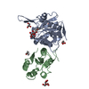
| ||||||||||||
|---|---|---|---|---|---|---|---|---|---|---|---|---|---|
| 1 |
| ||||||||||||
| Unit cell |
| ||||||||||||
| Components on special symmetry positions |
|
-Components
-Protein , 2 types, 2 molecules AB
| #1: Protein | Asparaginase Mass: 22265.480 Da / Num. of mol.: 1 / Fragment: UNP residues 1-182 Source method: isolated from a genetically manipulated source Source: (gene. exp.) Pyrococcus furiosus DSM 3638 (archaea) / Gene: PF2047, ph0066 / Plasmid: pET-28a / Production host:  Escherichia coli (E. coli) / Strain (production host): Rosetta (DE3) / References: UniProt: Q8TZE8, asparaginase Escherichia coli (E. coli) / Strain (production host): Rosetta (DE3) / References: UniProt: Q8TZE8, asparaginase |
|---|---|
| #2: Protein | Asparaginase Mass: 16075.715 Da / Num. of mol.: 1 / Fragment: UNP residues 202-326 Source method: isolated from a genetically manipulated source Source: (gene. exp.) Pyrococcus furiosus DSM 3638 (archaea) / Gene: PF2047, ph0066 / Plasmid: pET-28a / Production host: Escherichia coli (E. coli) / Strain (production host): Rosetta (DE3) / References: UniProt: Q8TZE8, asparaginase |
-Non-polymers , 4 types, 138 molecules 


| #3: Chemical | Citric acid Mass: 189.100 Da / Num. of mol.: 2 / Source method: obtained synthetically / Formula: C6H5O7 Mass: 189.100 Da / Num. of mol.: 2 / Source method: obtained synthetically / Formula: C6H5O7#4: Chemical | ChemComp-GOL / Glycerol Mass: 92.094 Da / Num. of mol.: 4 / Source method: obtained synthetically / Formula: C3H8O3 Mass: 92.094 Da / Num. of mol.: 4 / Source method: obtained synthetically / Formula: C3H8O3#5: Chemical | Isopropyl alcohol Mass: 60.095 Da / Num. of mol.: 2 / Source method: obtained synthetically / Formula: C3H8O / Comment: alkaloid*YM Mass: 60.095 Da / Num. of mol.: 2 / Source method: obtained synthetically / Formula: C3H8O / Comment: alkaloid*YM#6: Water | ChemComp-HOH / | WaterMass: 18.015 Da / Num. of mol.: 130 / Source method: isolated from a natural source / Formula: H2O |
|---|
-Experimental details
-Experiment
| Experiment | Method: X-RAY DIFFRACTION / Number of used crystals: 1 |
|---|
- Sample preparation
| Crystal | Density Matthews: 2.94 Å3/Da / Density % sol: 58.16 % |
|---|---|
| Crystal grow | Temperature: 293 K / Method: vapor diffusion, hanging drop / pH: 6 Details: 200MM SODIUM CITRATE TRIBASIC DIHYDRATE, 100MM SODIUM CACODYLATE, 30%(V/V) 2-PROPANOL, pH 6.0, VAPOR DIFFUSION, HANGING DROP, temperature 293K |
-Data collection
| Diffraction | Mean temperature: 100 K |
|---|---|
| Diffraction source | Source: ROTATING ANODE / Type: RIGAKU MICROMAX-007 HF / Wavelength: 1.5418 Å |
| Detector | Type: MAR scanner 345 mm plate / Detector: IMAGE PLATE / Date: Nov 14, 2013 / Details: Mirrors |
| Radiation | Monochromator: Mirrors / Protocol: SINGLE WAVELENGTH / Monochromatic (M) / Laue (L): M / Scattering type: x-ray |
| Radiation wavelength | Wavelength: 1.5418 Å / Relative weight: 1 |
| Reflection | Resolution: 2.049→50 Å / Num. obs: 29876 / % possible obs: 99.8 % / Observed criterion σ(F): 2 / Observed criterion σ(I): 2 / Redundancy: 14.5 % / Biso Wilson estimate: 37.28 Å2 / Rmerge(I) obs: 0.089 / Rsym value: 0.089 / Net I/σ(I): 38.8 |
| Reflection shell | Resolution: 2.05→2.09 Å / Redundancy: 9.2 % / Rmerge(I) obs: 0.79 / Mean I/σ(I) obs: 2.05 / Num. unique all: 1424 / Rsym value: 0.79 / % possible all: 97.5 |
- Processing
| Software |
| ||||||||||||||||||||||||||||||||||||||||||||||||||||||||||||||||||||||||||||||||||||
|---|---|---|---|---|---|---|---|---|---|---|---|---|---|---|---|---|---|---|---|---|---|---|---|---|---|---|---|---|---|---|---|---|---|---|---|---|---|---|---|---|---|---|---|---|---|---|---|---|---|---|---|---|---|---|---|---|---|---|---|---|---|---|---|---|---|---|---|---|---|---|---|---|---|---|---|---|---|---|---|---|---|---|---|---|---|
| Refinement | Method to determine structure: MOLECULAR REPLACEMENT Starting model: 4NJE Resolution: 2.049→33.37 Å / FOM work R set: 0.81 / SU ML: 0.24 / Isotropic thermal model: Isotropic / σ(F): 1.33 / Phase error: 24.54 / Stereochemistry target values: ML
| ||||||||||||||||||||||||||||||||||||||||||||||||||||||||||||||||||||||||||||||||||||
| Solvent computation | Shrinkage radii: 0.86 Å / VDW probe radii: 1.1 Å / Solvent model: FLAT BULK SOLVENT MODEL / Bsol: 59.572 Å2 / ksol: 0.4 e/Å3 | ||||||||||||||||||||||||||||||||||||||||||||||||||||||||||||||||||||||||||||||||||||
| Displacement parameters | Biso max: 100.69 Å2 / Biso mean: 41.44 Å2 / Biso min: 22.49 Å2
| ||||||||||||||||||||||||||||||||||||||||||||||||||||||||||||||||||||||||||||||||||||
| Refinement step | Cycle: LAST / Resolution: 2.049→33.37 Å
| ||||||||||||||||||||||||||||||||||||||||||||||||||||||||||||||||||||||||||||||||||||
| Refine LS restraints |
| ||||||||||||||||||||||||||||||||||||||||||||||||||||||||||||||||||||||||||||||||||||
| LS refinement shell | Refine-ID: X-RAY DIFFRACTION / Total num. of bins used: 11
|
