 Movie
Movie Controller


+ Open data

- Basic information
| Entry | Database: PDB / ID: 4r7x | ||||||
|---|---|---|---|---|---|---|---|
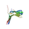
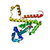
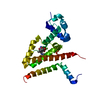
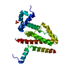
| Title | Crystal structure of N-lobe of human ARRDC3(1-180) | ||||||
 Components Components | Arrestin domain-containing protein 3 | ||||||
 Keywords Keywords |  PROTEIN BINDING / arrestin fold / GPCR downregulation / Beat 2 adrenergic receptor PROTEIN BINDING / arrestin fold / GPCR downregulation / Beat 2 adrenergic receptor | ||||||
| Function / homology |  Function and homology information Function and homology informationnegative regulation of heat generation / beta-3 adrenergic receptor binding / negative regulation of locomotion involved in locomotory behavior / positive regulation of hippo signaling / negative regulation of adenylate cyclase-activating adrenergic receptor signaling pathway / positive regulation of ubiquitin-protein transferase activity / heat generation / negative regulation of cold-induced thermogenesis / fat pad development / skin development ...negative regulation of heat generation / beta-3 adrenergic receptor binding / negative regulation of locomotion involved in locomotory behavior / positive regulation of hippo signaling / negative regulation of adenylate cyclase-activating adrenergic receptor signaling pathway / positive regulation of ubiquitin-protein transferase activity / heat generation / negative regulation of cold-induced thermogenesis / fat pad development / skin development / protein transport / lysosome / early endosome / endosome / plasma membrane / cytoplasmSimilarity search - Function | ||||||
| Biological species |  Homo sapiens (human) Homo sapiens (human) | ||||||
| Method | X-RAY DIFFRACTION / SYNCHROTRON / MOLECULAR REPLACEMENT / Resolution: 2.61 Å | ||||||
 Authors Authors | Qi, S. / Hurley, J. | ||||||
 Citation Citation | Journal: Protein Sci. / Year: 2014 Title: Insights into beta 2-adrenergic receptor binding from structures of the N-terminal lobe of ARRDC3. Authors: Qi, S. / O'Hayre, M. / Gutkind, J.S. / Hurley, J.H. | ||||||
| History |
|
- Structure visualization
| Structure viewer | Molecule: MolmilJmol/JSmol |
|---|
- Downloads & links
-Download
| PDBx/mmCIF format | 4r7x.cif.gz | 136.8 KB | Display | PDBx/mmCIF format |
|---|---|---|---|---|
| PDB format | pdb4r7x.ent.gz | 108.1 KB | Display | PDB format |
| PDBx/mmJSON format | 4r7x.json.gz | Tree view | PDBx/mmJSON format | |
| Others |  Other downloads Other downloads |
-Validation report
| Arichive directory | https://data.pdbj.org/pub/pdb/validation_reports/r7/4r7xftp://data.pdbj.org/pub/pdb/validation_reports/r7/4r7x | HTTPS FTP |
|---|
-Related structure data


-Links
 PDBj
PDBj- Assembly
| Deposited unit | 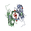
| ||||||||
|---|---|---|---|---|---|---|---|---|---|
| 1 | 
| ||||||||
| 2 | 
| ||||||||
| Unit cell |
| ||||||||
| Components on special symmetry positions |
|
-Components
| #1: Protein | Mass: 20891.338 Da / Num. of mol.: 2 / Fragment: UNP residues 1-180 Source method: isolated from a genetically manipulated source Source: (gene. exp.) Homo sapiens (human) / Gene: ARRDC3, KIAA1376 / Production host:  Escherichia coli (E. coli) / References: UniProt: Q96B67 Escherichia coli (E. coli) / References: UniProt: Q96B67#2: Chemical | ChemComp-PO4 / Phosphate  Mass: 94.971 Da / Num. of mol.: 11 / Source method: obtained synthetically / Formula: PO4 Mass: 94.971 Da / Num. of mol.: 11 / Source method: obtained synthetically / Formula: PO4#3: Water | ChemComp-HOH / | Water Mass: 18.015 Da / Num. of mol.: 13 / Source method: isolated from a natural source / Formula: H2O Mass: 18.015 Da / Num. of mol.: 13 / Source method: isolated from a natural source / Formula: H2O |
|---|
-Experimental details
-Experiment
| Experiment | Method: X-RAY DIFFRACTION / Number of used crystals: 1 |
|---|
- Sample preparation
| Crystal | Density Matthews: 4.11 Å3/Da / Density % sol: 70.04 % |
|---|---|
| Crystal grow | Temperature: 294 K / Method: vapor diffusion, hanging drop / pH: 8 Details: 0.75M NH4H2PO4, 0.1M Sodium citrate tribasic dehydrate, 3.0% (W/V) 6-Aminohexanoic acid, pH 8.0, VAPOR DIFFUSION, HANGING DROP, temperature 294K |
-Data collection
| Diffraction | Mean temperature: 100 K |
|---|---|
| Diffraction source | Source: SYNCHROTRON / Site: APS  / Beamline: 22-ID / Wavelength: 1 Å / Beamline: 22-ID / Wavelength: 1 Å |
| Detector | Type: RAYONIX MX300HS / Detector: CCD / Date: Jun 27, 2013 |
| Radiation | Monochromator: Si(111) / Protocol: SINGLE WAVELENGTH / Monochromatic (M) / Laue (L): M / Scattering type: x-ray |
| Radiation wavelength | Wavelength: 1 Å / Relative weight: 1 |
| Reflection | Resolution: 2.61→50 Å / Num. all: 21676 / Num. obs: 20902 / % possible obs: 96.4 % / Observed criterion σ(F): 1 / Observed criterion σ(I): 1 |
| Reflection shell | Resolution: 2.61→2.74 Å / % possible all: 99.9 |
- Processing
| Software |
| |||||||||||||||||||||||||||||||||||||||||||||||||||||||||||||||
|---|---|---|---|---|---|---|---|---|---|---|---|---|---|---|---|---|---|---|---|---|---|---|---|---|---|---|---|---|---|---|---|---|---|---|---|---|---|---|---|---|---|---|---|---|---|---|---|---|---|---|---|---|---|---|---|---|---|---|---|---|---|---|---|---|
| Refinement | Method to determine structure: MOLECULAR REPLACEMENT / Resolution: 2.61→32.026 Å / SU ML: 0.47 / σ(F): 1.33 / Phase error: 29.27 / Stereochemistry target values: ML
| |||||||||||||||||||||||||||||||||||||||||||||||||||||||||||||||
| Solvent computation | Shrinkage radii: 0.9 Å / VDW probe radii: 1.11 Å / Solvent model: FLAT BULK SOLVENT MODEL | |||||||||||||||||||||||||||||||||||||||||||||||||||||||||||||||
| Refinement step | Cycle: LAST / Resolution: 2.61→32.026 Å
| |||||||||||||||||||||||||||||||||||||||||||||||||||||||||||||||
| Refine LS restraints |
| |||||||||||||||||||||||||||||||||||||||||||||||||||||||||||||||
| LS refinement shell |
| |||||||||||||||||||||||||||||||||||||||||||||||||||||||||||||||
| Refinement TLS params. | Method: refined / Origin x: -2.7002 Å / Origin y: 32.3354 Å / Origin z: 14.8749 Å
| |||||||||||||||||||||||||||||||||||||||||||||||||||||||||||||||
| Refinement TLS group |
|