 Movie
Movie Controller

[English] 日本語
 Yorodumi
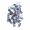
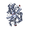
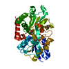
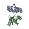
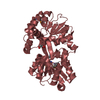
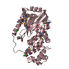
- PDB-4r72: Structure of the periplasmic binding protein AfuA from Actinobaci... -
+ Open data

- Basic information
| Entry | Database: PDB / ID: 4r72 | ||||||
|---|---|---|---|---|---|---|---|
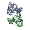
| Title | Structure of the periplasmic binding protein AfuA from Actinobacillus pleuropneumoniae (apo form) | ||||||
 Components Components | ABC-type Fe3+ transport system, periplasmic component | ||||||
 Keywords Keywords |  TRANSPORT PROTEIN / ABC transporter / sugar transporter / glucose-6-phosphate / fructose-6-phosphate / sedoheptulose-7-phosphate TRANSPORT PROTEIN / ABC transporter / sugar transporter / glucose-6-phosphate / fructose-6-phosphate / sedoheptulose-7-phosphate | ||||||
| Function / homology | Bacterial extracellular solute-binding protein / Ferric binding protein / Periplasmic binding protein-like II / D-Maltodextrin-Binding Protein; domain 2 / 3-Layer(aba) Sandwich / metal ion binding / Alpha Beta / DI(HYDROXYETHYL)ETHER / ABC-type Fe3+ transport system, periplasmic component Function and homology information Function and homology information | ||||||
| Biological species |  Actinobacillus pleuropneumoniae (bacteria) Actinobacillus pleuropneumoniae (bacteria) | ||||||
| Method | X-RAY DIFFRACTION / SYNCHROTRON / MOLECULAR REPLACEMENT / Resolution: 1.6 Å | ||||||
 Authors Authors | Sit, B. / Calmettes, C. / Moraes, T.F. | ||||||
 Citation Citation | Journal: Plos Pathog. / Year: 2015 Title: Active Transport of Phosphorylated Carbohydrates Promotes Intestinal Colonization and Transmission of a Bacterial Pathogen. Authors: Sit, B. / Crowley, S.M. / Bhullar, K. / Lai, C.C. / Tang, C. / Hooda, Y. / Calmettes, C. / Khambati, H. / Ma, C. / Brumell, J.H. / Schryvers, A.B. / Vallance, B.A. / Moraes, T.F. | ||||||
| History |
|
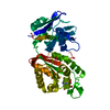
- Structure visualization
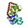
| Structure viewer | Molecule: MolmilJmol/JSmol |
|---|
- Downloads & links
-Download
| PDBx/mmCIF format | 4r72.cif.gz | 85.3 KB | Display | PDBx/mmCIF format |
|---|---|---|---|---|
| PDB format | pdb4r72.ent.gz | 66.6 KB | Display | PDB format |
| PDBx/mmJSON format | 4r72.json.gz | Tree view | PDBx/mmJSON format | |
| Others |  Other downloads Other downloads |
-Validation report
| Arichive directory | https://data.pdbj.org/pub/pdb/validation_reports/r7/4r72ftp://data.pdbj.org/pub/pdb/validation_reports/r7/4r72 | HTTPS FTP |
|---|
-Related structure data


-Links
 PDBj
PDBj- Assembly
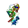
| Deposited unit | 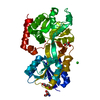
| ||||||||||||||||||
|---|---|---|---|---|---|---|---|---|---|---|---|---|---|---|---|---|---|---|---|
| 1 |
| ||||||||||||||||||
| Unit cell |
| ||||||||||||||||||
| Components on special symmetry positions |
|
-Components
-Protein , 1 types, 1 molecules A
| #1: Protein | Mass: 35391.980 Da / Num. of mol.: 1 / Fragment: UNP residues 28-346 Source method: isolated from a genetically manipulated source Source: (gene. exp.) Actinobacillus pleuropneumoniae (bacteria)Gene: afuA, APL_1446 / Production host: Escherichia coli (E. coli) / References: UniProt: A3N294 |
|---|
-Non-polymers , 5 types, 433 molecules 


| #2: Chemical | ChemComp-GOL / Glycerol Mass: 92.094 Da / Num. of mol.: 5 / Source method: obtained synthetically / Formula: C3H8O3 Mass: 92.094 Da / Num. of mol.: 5 / Source method: obtained synthetically / Formula: C3H8O3#3: Chemical | ChemComp-PEG / | Diethylene glycol Mass: 106.120 Da / Num. of mol.: 1 / Source method: obtained synthetically / Formula: C4H10O3 Mass: 106.120 Da / Num. of mol.: 1 / Source method: obtained synthetically / Formula: C4H10O3#4: Chemical | ChemComp-TRS / | Tris Mass: 122.143 Da / Num. of mol.: 1 / Source method: obtained synthetically / Formula: C4H12NO3 / Comment: pH buffer*YM Mass: 122.143 Da / Num. of mol.: 1 / Source method: obtained synthetically / Formula: C4H12NO3 / Comment: pH buffer*YM#5: Chemical | ChemComp-CL / | Chloride Mass: 35.453 Da / Num. of mol.: 1 / Source method: obtained synthetically / Formula: Cl Mass: 35.453 Da / Num. of mol.: 1 / Source method: obtained synthetically / Formula: Cl#6: Water | ChemComp-HOH / | WaterMass: 18.015 Da / Num. of mol.: 425 / Source method: isolated from a natural source / Formula: H2O |
|---|
-Experimental details
-Experiment
| Experiment | Method: X-RAY DIFFRACTION / Number of used crystals: 1 |
|---|
- Sample preparation
| Crystal | Density Matthews: 2.08 Å3/Da / Density % sol: 41 % |
|---|---|
| Crystal grow | Temperature: 298 K / Method: vapor diffusion, sitting drop / pH: 6 Details: 0.2 M magnesium chloride, 0.1 M MES/NaOH, pH 6.0, 27.5% PEG3350, 20% glycerol, VAPOR DIFFUSION, SITTING DROP, temperature 298K |
-Data collection
| Diffraction | Mean temperature: 100 K | |||||||||||||||||||||||||||||||||||||||||||||||||||
|---|---|---|---|---|---|---|---|---|---|---|---|---|---|---|---|---|---|---|---|---|---|---|---|---|---|---|---|---|---|---|---|---|---|---|---|---|---|---|---|---|---|---|---|---|---|---|---|---|---|---|---|---|
| Diffraction source | Source: SYNCHROTRON / Site: CLSI  / Beamline: 08ID-1 / Wavelength: 0.9795 Å / Beamline: 08ID-1 / Wavelength: 0.9795 Å | |||||||||||||||||||||||||||||||||||||||||||||||||||
| Detector | Type: RAYONIX MX-300 / Detector: CCD | |||||||||||||||||||||||||||||||||||||||||||||||||||
| Radiation | Monochromator: ACCEL/BRUKER double crystal Si(111) / Protocol: SINGLE WAVELENGTH / Monochromatic (M) / Laue (L): M / Scattering type: x-ray | |||||||||||||||||||||||||||||||||||||||||||||||||||
| Radiation wavelength | Wavelength: 0.9795 Å / Relative weight: 1 | |||||||||||||||||||||||||||||||||||||||||||||||||||
| Reflection | Resolution: 1.6→47.095 Å / Num. obs: 38225 / Biso Wilson estimate: 18.12 Å2 | |||||||||||||||||||||||||||||||||||||||||||||||||||
| Reflection shell |
|
- Processing
| Software |
| |||||||||||||||||||||||||||||||||||||||||||||||||||||||||||||||||||||||||||||||||||||||||||||||||||||||||
|---|---|---|---|---|---|---|---|---|---|---|---|---|---|---|---|---|---|---|---|---|---|---|---|---|---|---|---|---|---|---|---|---|---|---|---|---|---|---|---|---|---|---|---|---|---|---|---|---|---|---|---|---|---|---|---|---|---|---|---|---|---|---|---|---|---|---|---|---|---|---|---|---|---|---|---|---|---|---|---|---|---|---|---|---|---|---|---|---|---|---|---|---|---|---|---|---|---|---|---|---|---|---|---|---|---|---|
| Refinement | Method to determine structure: MOLECULAR REPLACEMENT / Resolution: 1.6→47.095 Å / SU ML: 0.14 / σ(F): 1.36 / Phase error: 19.11 / Stereochemistry target values: ML
| |||||||||||||||||||||||||||||||||||||||||||||||||||||||||||||||||||||||||||||||||||||||||||||||||||||||||
| Solvent computation | Shrinkage radii: 0.9 Å / VDW probe radii: 1.11 Å / Solvent model: FLAT BULK SOLVENT MODEL | |||||||||||||||||||||||||||||||||||||||||||||||||||||||||||||||||||||||||||||||||||||||||||||||||||||||||
| Displacement parameters | Biso max: 79.77 Å2 / Biso mean: 23.093 Å2 / Biso min: 9.22 Å2 | |||||||||||||||||||||||||||||||||||||||||||||||||||||||||||||||||||||||||||||||||||||||||||||||||||||||||
| Refinement step | Cycle: LAST / Resolution: 1.6→47.095 Å
| |||||||||||||||||||||||||||||||||||||||||||||||||||||||||||||||||||||||||||||||||||||||||||||||||||||||||
| Refine LS restraints |
| |||||||||||||||||||||||||||||||||||||||||||||||||||||||||||||||||||||||||||||||||||||||||||||||||||||||||
| LS refinement shell | Refine-ID: X-RAY DIFFRACTION / Total num. of bins used: 14
|
