 Movie
Movie Controller

[English] 日本語
 Yorodumi
- PDB-4r6o: Jacalin-carbohydrate interactions. Distortion of the ligand as a ... -
+ Open data

- Basic information
| Entry | Database: PDB / ID: 4r6o | ||||||
|---|---|---|---|---|---|---|---|
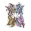
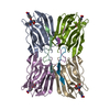
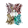
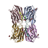
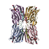
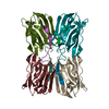
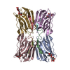
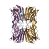
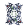
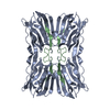
| Title | Jacalin-carbohydrate interactions. Distortion of the ligand as a determinant of affinity. | ||||||
 Components Components |
| ||||||
 Keywords Keywords | SUGAR BINDING PROTEIN / Galactose specific lectin / beta-prism I fold / post translational proteolysis / T-antigen binding protein /  Plant lectins / Galactose Plant lectins / Galactose | ||||||
| Function / homology |  Function and homology information Function and homology information | ||||||
| Biological species |   Artocarpus integer (campedak) Artocarpus integer (campedak) | ||||||
| Method | X-RAY DIFFRACTION / SYNCHROTRON / MOLECULAR REPLACEMENT / Resolution: 1.6 Å | ||||||
 Authors Authors | Abhinav, K.V. / Sharma, K. / Swaminathan, C.P. / Surolia, A. / Vijayan, M. | ||||||
 Citation Citation | Journal: Acta Crystallogr.,Sect.D / Year: 2015 Title: Jacalin-carbohydrate interactions: distortion of the ligand molecule as a determinant of affinity. Authors: Abhinav, K.V. / Sharma, K. / Swaminathan, C.P. / Surolia, A. / Vijayan, M. #1: Journal: Nat.Struct.Biol. / Year: 1996Title: A novel mode of carbohydrate recognition in jacalin, a Moraceae plant lectin with a beta-prism fold. Authors: Sankaranarayanan, R. / Sekar, K. / Banerjee, R. / Sharma, V. / Surolia, A. / Vijayan, M. #2: Journal: J.Mol.Biol. / Year: 2002Title: Crystal structure of the jacalin-T-antigen complex and a comparative study of lectin-T-antigen complexes. Authors: Jeyaprakash, A.A. / Geetha Rani, P. / Banuprakash Reddy, G. / Banumathi, S. / Betzel, C. / Sekar, K. / Surolia, A. / Vijayan, M. #3: Journal: J.Mol.Biol. / Year: 2003Title: Structural basis of the carbohydrate specificities of jacalin: an X-ray and modeling study. Authors: Jeyaprakash, A.A. / Katiyar, S. / Swaminathan, C.P. / Sekar, K. / Surolia, A. / Vijayan, M. #4: Journal: J.Mol.Biol. / Year: 2005Title: Structural basis for the energetics of jacalin-sugar interactions: promiscuity versus specificity. Authors: Arockia Jeyaprakash, A. / Jayashree, G. / Mahanta, S.K. / Swaminathan, C.P. / Sekar, K. / Surolia, A. / Vijayan, M. #5: Journal: Biochem.J. / Year: 2002Title: Structural basis for the unusual carbohydrate-binding specificity of jacalin towards galactose and mannose. Authors: Bourne, Y. / Astoul, C.H. / Zamboni, V. / Peumans, W.J. / Menu-Bouaouiche, L. / Van Damme, E.J. / Barre, A. / Rouge, P. | ||||||
| History |
|
- Structure visualization
| Structure viewer | Molecule: MolmilJmol/JSmol |
|---|
- Downloads & links
-Download
| PDBx/mmCIF format | 4r6o.cif.gz | 144.1 KB | Display | PDBx/mmCIF format |
|---|---|---|---|---|
| PDB format | pdb4r6o.ent.gz | 112.6 KB | Display | PDB format |
| PDBx/mmJSON format | 4r6o.json.gz | Tree view | PDBx/mmJSON format | |
| Others |  Other downloads Other downloads |
-Validation report
| Arichive directory | https://data.pdbj.org/pub/pdb/validation_reports/r6/4r6oftp://data.pdbj.org/pub/pdb/validation_reports/r6/4r6o | HTTPS FTP |
|---|
-Related structure data
| Related structure data | 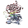 4r6nC 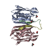 4r6pC 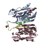 4r6qC 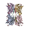 4r6rC  1ku8S S: Starting model for refinement C: citing same article ( |
|---|---|
| Similar structure data |


-Links
 PDBj
PDBj
- Assembly
| Deposited unit | 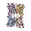
| ||||||||
|---|---|---|---|---|---|---|---|---|---|
| 1 |
| ||||||||
| 2 | 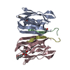
| ||||||||
| 3 | 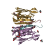
| ||||||||
| Unit cell |
|
-Components
-Protein / Protein/peptide / Sugars , 3 types, 12 molecules ACEGBDFH
| #1: Protein | Mass: 14673.479 Da / Num. of mol.: 4 / Fragment: UNP RESIDUES 2-20 / Source method: isolated from a natural source / Source: (natural) Artocarpus integer (campedak) / References: UniProt: P18670#2: Protein/peptide | Mass: 1944.170 Da / Num. of mol.: 4 / Source method: isolated from a natural source / Source: (natural) Artocarpus integer (campedak) / References: UniProt: P18673#4: Sugar | ChemComp-GLA / Galactose Type: D-saccharide, alpha linking / Mass: 180.156 Da / Num. of mol.: 4 Type: D-saccharide, alpha linking / Mass: 180.156 Da / Num. of mol.: 4Source method: isolated from a genetically manipulated source Formula: C6H12O6 |
|---|
-Non-polymers , 4 types, 690 molecules 


| #3: Chemical | ChemComp-ZZ1 /  Mass: 160.169 Da / Num. of mol.: 4 / Source method: obtained synthetically / Formula: C10H8O2 Mass: 160.169 Da / Num. of mol.: 4 / Source method: obtained synthetically / Formula: C10H8O2#5: Chemical | ChemComp-IPA / Isopropyl alcohol Mass: 60.095 Da / Num. of mol.: 11 / Source method: obtained synthetically / Formula: C3H8O / Comment: alkaloid*YM Mass: 60.095 Da / Num. of mol.: 11 / Source method: obtained synthetically / Formula: C3H8O / Comment: alkaloid*YM#6: Chemical | Ethylene glycol Mass: 62.068 Da / Num. of mol.: 2 / Source method: obtained synthetically / Formula: C2H6O2 Mass: 62.068 Da / Num. of mol.: 2 / Source method: obtained synthetically / Formula: C2H6O2#7: Water | ChemComp-HOH / | WaterMass: 18.015 Da / Num. of mol.: 673 / Source method: isolated from a natural source / Formula: H2O |
|---|
-Experimental details
-Experiment
| Experiment | Method: X-RAY DIFFRACTION / Number of used crystals: 1 |
|---|
- Sample preparation
| Crystal | Density Matthews: 2.2 Å3/Da / Density % sol: 43.98 % |
|---|---|
| Crystal grow | Temperature: 298 K / Method: vapor diffusion, hanging drop / pH: 7.4 Details: 15% PEG 8000, 0.1M HEPES, pH 7.4, 10% (v/v) isopropanol, VAPOR DIFFUSION, HANGING DROP, temperature 298K |
-Data collection
| Diffraction | Mean temperature: 100 K | ||||||||||||||||||||||||||||
|---|---|---|---|---|---|---|---|---|---|---|---|---|---|---|---|---|---|---|---|---|---|---|---|---|---|---|---|---|---|
| Diffraction source | Source: SYNCHROTRON / Site: ESRF  / Beamline: BM14 / Wavelength: 0.98 Å / Beamline: BM14 / Wavelength: 0.98 Å | ||||||||||||||||||||||||||||
| Detector | Type: MARMOSAIC 225 mm CCD / Detector: CCD / Date: Oct 21, 2013 / Details: Bent collimating mirror and toroid | ||||||||||||||||||||||||||||
| Radiation | Monochromator: Si(111) monochromator. / Protocol: SINGLE WAVELENGTH / Monochromatic (M) / Laue (L): M / Scattering type: x-ray | ||||||||||||||||||||||||||||
| Radiation wavelength | Wavelength: 0.98 Å / Relative weight: 1 | ||||||||||||||||||||||||||||
| Reflection | Resolution: 1.55→60.62 Å / Num. all: 83181 / Num. obs: 83181 / % possible obs: 99.9 % / Observed criterion σ(F): 0 / Observed criterion σ(I): 0 / Redundancy: 4.2 % / Rsym value: 0.089 / Net I/σ(I): 8.2 | ||||||||||||||||||||||||||||
| Reflection shell |
|
- Processing
| Software |
| |||||||||||||||||||||||||||||||||||||||||||||
|---|---|---|---|---|---|---|---|---|---|---|---|---|---|---|---|---|---|---|---|---|---|---|---|---|---|---|---|---|---|---|---|---|---|---|---|---|---|---|---|---|---|---|---|---|---|---|
| Refinement | Method to determine structure: MOLECULAR REPLACEMENT Starting model: 1KU8 Resolution: 1.6→28.05 Å / Cor.coef. Fo:Fc: 0.966 / Cor.coef. Fo:Fc free: 0.948 / SU B: 2.034 / SU ML: 0.071 / Cross valid method: THROUGHOUT / σ(F): 0 / σ(I): 0 / ESU R: 0.096 / ESU R Free: 0.098 / Stereochemistry target values: MAXIMUM LIKELIHOOD / Details: HYDROGENS HAVE BEEN USED IF PRESENT IN THE INPUT
| |||||||||||||||||||||||||||||||||||||||||||||
| Solvent computation | Ion probe radii: 0.8 Å / Shrinkage radii: 0.8 Å / VDW probe radii: 1.2 Å / Solvent model: MASK | |||||||||||||||||||||||||||||||||||||||||||||
| Displacement parameters | Biso mean: 22.066 Å2
| |||||||||||||||||||||||||||||||||||||||||||||
| Refinement step | Cycle: LAST / Resolution: 1.6→28.05 Å
| |||||||||||||||||||||||||||||||||||||||||||||
| Refine LS restraints |
| |||||||||||||||||||||||||||||||||||||||||||||
| LS refinement shell | Resolution: 1.6→1.642 Å / Total num. of bins used: 20
|
