 Movie
Movie Controller

[English] 日本語
 Yorodumi
- PDB-4r1o: Crystal Structure of Thermophilic Geobacillus kaustophilus L-Arab... -
+ Open data

- Basic information
| Entry | Database: PDB / ID: 4r1o | ||||||
|---|---|---|---|---|---|---|---|
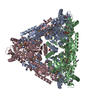
| Title | Crystal Structure of Thermophilic Geobacillus kaustophilus L-Arabinose isomerase | ||||||
 Components Components | L-arabinose isomerase | ||||||
 Keywords Keywords | ISOMERASE / Hexamer / Thermophile / thermostable / AI fold | ||||||
| Function / homology |  Function and homology informationL-arabinose isomerase / L-arabinose isomerase activity / L-arabinose catabolic process to xylulose 5-phosphate / manganese ion binding / cytosol Function and homology informationL-arabinose isomerase / L-arabinose isomerase activity / L-arabinose catabolic process to xylulose 5-phosphate / manganese ion binding / cytosolSimilarity search - Function | ||||||
| Biological species |  Geobacillus kaustophilus HTA426 (bacteria) Geobacillus kaustophilus HTA426 (bacteria) | ||||||
| Method | X-RAY DIFFRACTION / SYNCHROTRON / MOLECULAR REPLACEMENT / Resolution: 2.401 Å | ||||||
 Authors Authors | Choi, J.M. / Lee, Y.J. / Lee, D.W. / Lee, S.H. | ||||||
 Citation Citation | Journal: to be published Title: Crystal Structure of Thermophilic apo L-Arabinose Isomerase from Geobacillus kaustophilus Authors: Choi, J.M. / Lee, Y.J. / Lee, D.W. / Lee, S.H. | ||||||
| History |
|
- Structure visualization
| Structure viewer | Molecule: MolmilJmol/JSmol |
|---|
- Downloads & links
-Download
| PDBx/mmCIF format | 4r1o.cif.gz | 594.7 KB | Display | PDBx/mmCIF format |
|---|---|---|---|---|
| PDB format | pdb4r1o.ent.gz | 492.5 KB | Display | PDB format |
| PDBx/mmJSON format | 4r1o.json.gz | Tree view | PDBx/mmJSON format | |
| Others |  Other downloads Other downloads |
-Validation report
| Arichive directory | https://data.pdbj.org/pub/pdb/validation_reports/r1/4r1oftp://data.pdbj.org/pub/pdb/validation_reports/r1/4r1o | HTTPS FTP |
|---|
-Related structure data
| Related structure data | |
|---|---|
| Similar structure data |


-Links
 PDBj
PDBj- Assembly
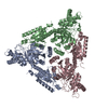
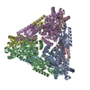
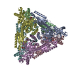
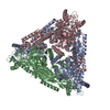
| Deposited unit | 
| ||||||||
|---|---|---|---|---|---|---|---|---|---|
| 1 |
| ||||||||
| Unit cell |
|
-Components
| #1: Protein | Mass: 56304.891 Da / Num. of mol.: 6 Source method: isolated from a genetically manipulated source Source: (gene. exp.) Geobacillus kaustophilus HTA426 (bacteria)Gene: araA, GK1904 / Plasmid: pET28a(+)-GKAI / Production host: Escherichia coli BL21(DE3) (bacteria) / References: UniProt: Q5KYP7, L-arabinose isomerase#2: Water | ChemComp-HOH / | Water Mass: 18.015 Da / Num. of mol.: 1182 / Source method: isolated from a natural source / Formula: H2O Mass: 18.015 Da / Num. of mol.: 1182 / Source method: isolated from a natural source / Formula: H2OSequence details | THIS RESIDUE IS BASED ON YP_147757 OF GENBANK. | |
|---|
-Experimental details
-Experiment
| Experiment | Method: X-RAY DIFFRACTION / Number of used crystals: 1 |
|---|
- Sample preparation
| Crystal | Density Matthews: 2.63 Å3/Da / Density % sol: 53.22 % |
|---|---|
| Crystal grow | Temperature: 293 K / Method: vapor diffusion, hanging drop / pH: 7.2 Details: 1.8M Na-K phosphate, pH 7.2, VAPOR DIFFUSION, HANGING DROP, temperature 293K |
-Data collection
| Diffraction | Mean temperature: 100 K |
|---|---|
| Diffraction source | Source: SYNCHROTRON / Site: PAL/PLS  / Beamline: 7A (6B, 6C1) / Wavelength: 1.0015 Å / Beamline: 7A (6B, 6C1) / Wavelength: 1.0015 Å |
| Detector | Type: ADSC QUANTUM 270 / Detector: CCD / Date: Nov 16, 2013 |
| Radiation | Monochromator: DCM Si(111) Crystal / Protocol: SINGLE WAVELENGTH / Monochromatic (M) / Laue (L): M / Scattering type: x-ray |
| Radiation wavelength | Wavelength: 1.0015 Å / Relative weight: 1 |
| Reflection | Resolution: 2.4→50 Å / Num. all: 133329 / Num. obs: 133329 / % possible obs: 96.2 % / Observed criterion σ(F): 0 / Observed criterion σ(I): -3 / Redundancy: 7.4 % / Biso Wilson estimate: 25.61 Å2 / Rmerge(I) obs: 0.115 / Rsym value: 0.115 / Net I/σ(I): 14.091 |
| Reflection shell | Resolution: 2.4→2.44 Å / Redundancy: 7.1 % / Rmerge(I) obs: 0.458 / Mean I/σ(I) obs: 3.746 / Num. unique all: 6024 / Rsym value: 0.458 / % possible all: 94.1 |
- Processing
| Software |
| |||||||||||||||||||||||||||||||||||||||||||||||||||||||||||||||||||||||||||||||||||||||||||||||||||||||||||||||||||||||||||||||||||||||||||||||||||||||||||||||||||||||||||||||||||||||||||||||||||||||||||||||||||||||||
|---|---|---|---|---|---|---|---|---|---|---|---|---|---|---|---|---|---|---|---|---|---|---|---|---|---|---|---|---|---|---|---|---|---|---|---|---|---|---|---|---|---|---|---|---|---|---|---|---|---|---|---|---|---|---|---|---|---|---|---|---|---|---|---|---|---|---|---|---|---|---|---|---|---|---|---|---|---|---|---|---|---|---|---|---|---|---|---|---|---|---|---|---|---|---|---|---|---|---|---|---|---|---|---|---|---|---|---|---|---|---|---|---|---|---|---|---|---|---|---|---|---|---|---|---|---|---|---|---|---|---|---|---|---|---|---|---|---|---|---|---|---|---|---|---|---|---|---|---|---|---|---|---|---|---|---|---|---|---|---|---|---|---|---|---|---|---|---|---|---|---|---|---|---|---|---|---|---|---|---|---|---|---|---|---|---|---|---|---|---|---|---|---|---|---|---|---|---|---|---|---|---|---|---|---|---|---|---|---|---|---|---|---|---|---|---|---|---|---|
| Refinement | Method to determine structure: MOLECULAR REPLACEMENT Starting model: GKAI with MN Resolution: 2.401→45.323 Å / FOM work R set: 0.8468 / SU ML: 0.27 / σ(F): 1.36 / Phase error: 22.26 / Stereochemistry target values: ML
| |||||||||||||||||||||||||||||||||||||||||||||||||||||||||||||||||||||||||||||||||||||||||||||||||||||||||||||||||||||||||||||||||||||||||||||||||||||||||||||||||||||||||||||||||||||||||||||||||||||||||||||||||||||||||
| Solvent computation | Shrinkage radii: 0.9 Å / VDW probe radii: 1.11 Å / Solvent model: FLAT BULK SOLVENT MODEL | |||||||||||||||||||||||||||||||||||||||||||||||||||||||||||||||||||||||||||||||||||||||||||||||||||||||||||||||||||||||||||||||||||||||||||||||||||||||||||||||||||||||||||||||||||||||||||||||||||||||||||||||||||||||||
| Displacement parameters | Biso max: 84.6 Å2 / Biso mean: 17.04 Å2 / Biso min: 1.02 Å2 | |||||||||||||||||||||||||||||||||||||||||||||||||||||||||||||||||||||||||||||||||||||||||||||||||||||||||||||||||||||||||||||||||||||||||||||||||||||||||||||||||||||||||||||||||||||||||||||||||||||||||||||||||||||||||
| Refinement step | Cycle: LAST / Resolution: 2.401→45.323 Å
| |||||||||||||||||||||||||||||||||||||||||||||||||||||||||||||||||||||||||||||||||||||||||||||||||||||||||||||||||||||||||||||||||||||||||||||||||||||||||||||||||||||||||||||||||||||||||||||||||||||||||||||||||||||||||
| Refine LS restraints |
| |||||||||||||||||||||||||||||||||||||||||||||||||||||||||||||||||||||||||||||||||||||||||||||||||||||||||||||||||||||||||||||||||||||||||||||||||||||||||||||||||||||||||||||||||||||||||||||||||||||||||||||||||||||||||
| LS refinement shell | Refine-ID: X-RAY DIFFRACTION / Total num. of bins used: 30
|
