 Movie
Movie Controller

[English] 日本語
 Yorodumi
- PDB-4r1f: Re-refined Human DNA topoisomerase IIa (ATPase and transducer dom... -
+ Open data

- Basic information
| Entry | Database: PDB / ID: 4r1f | ||||||
|---|---|---|---|---|---|---|---|
| Title | Re-refined Human DNA topoisomerase IIa (ATPase and transducer domains) in complex with ADP and SO4 | ||||||
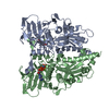
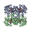
 Components Components | DNA topoisomerase 2-alpha | ||||||
 Keywords Keywords |  ISOMERASE / Human topoisomerase IIA ISOMERASE / Human topoisomerase IIA | ||||||
| Function / homology |  Function and homology information Function and homology informationnegative regulation of DNA duplex unwinding / positive regulation of single stranded viral RNA replication via double stranded DNA intermediate / sister chromatid segregation / apoptotic chromosome condensation / DNA topoisomerase type II (double strand cut, ATP-hydrolyzing) complex / resolution of meiotic recombination intermediates / female meiotic nuclear division / embryonic cleavage / DNA ligation / Transcription of E2F targets under negative control by DREAM complex ...negative regulation of DNA duplex unwinding / positive regulation of single stranded viral RNA replication via double stranded DNA intermediate / sister chromatid segregation / apoptotic chromosome condensation / DNA topoisomerase type II (double strand cut, ATP-hydrolyzing) complex / resolution of meiotic recombination intermediates / female meiotic nuclear division / embryonic cleavage / DNA ligation / Transcription of E2F targets under negative control by DREAM complex / DNA topoisomerase type II (double strand cut, ATP-hydrolyzing) activity / DNA topoisomerase (ATP-hydrolysing) / DNA binding, bending / DNA topological change / SUMOylation of DNA replication proteins / chromosome, centromeric region / ATP-dependent activity, acting on DNA / hematopoietic progenitor cell differentiation / condensed chromosome / protein kinase C binding / ubiquitin binding / male germ cell nucleus / chromosome segregation / regulation of circadian rhythm / rhythmic process / ribonucleoprotein complex / positive regulation of apoptotic process / protein heterodimerization activity / DNA damage response / chromatin binding / nucleolus / magnesium ion binding / protein homodimerization activity / positive regulation of transcription by RNA polymerase II / protein-containing complex / DNA binding / RNA binding / nucleoplasm / ATP binding / nucleus / cytoplasmSimilarity search - Function | ||||||
| Biological species |  Homo sapiens (human) Homo sapiens (human) | ||||||
| Method | X-RAY DIFFRACTION / MOLECULAR REPLACEMENT / Resolution: 2.51 Å | ||||||
 Authors Authors | Stanger, F.V. / Schirmer, T. | ||||||
 Citation Citation | Journal: Plos One / Year: 2014 Title: Structure of the N-Terminal Gyrase B Fragment in Complex with ADPPi Reveals Rigid-Body Motion Induced by ATP Hydrolysis Authors: Stanger, F.V. / Dehio, C. / Schirmer, T. #1: Journal: J.Biol.Chem. / Year: 2005Title: NUCLEOTIDE-DEPENDENT DOMAIN MOVEMENT in THE ATPASE DOMAIN of A HUMAN TYPE IIA DNA TOPOISOMERASE Authors: Wei, H. / Ruthenburg, A.J. / Bechis, S.K. / Verdine, G.L. | ||||||
| History |
| ||||||
| Remark 0 | THIS ENTRY 4R1F REFLECTS AN ALTERNATIVE MODELING OF THE ORIGINAL STRUCTURAL DATA (R1ZXNSF) ...THIS ENTRY 4R1F REFLECTS AN ALTERNATIVE MODELING OF THE ORIGINAL STRUCTURAL DATA (R1ZXNSF) DETERMINED BY AUTHORS OF THE PDB ENTRY 1ZXN: AUTHOR INITIALS, AUTHOR LAST NAME |
- Structure visualization
| Structure viewer | Molecule: MolmilJmol/JSmol |
|---|
- Downloads & links
-Download
| PDBx/mmCIF format | 4r1f.cif.gz | 315.4 KB | Display | PDBx/mmCIF format |
|---|---|---|---|---|
| PDB format | pdb4r1f.ent.gz | 255.4 KB | Display | PDB format |
| PDBx/mmJSON format | 4r1f.json.gz | Tree view | PDBx/mmJSON format | |
| Others |  Other downloads Other downloads |
-Validation report
| Arichive directory | https://data.pdbj.org/pub/pdb/validation_reports/r1/4r1fftp://data.pdbj.org/pub/pdb/validation_reports/r1/4r1f | HTTPS FTP |
|---|
-Related structure data
| Related structure data | 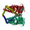 4prvC 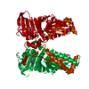 4prxC 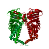 4pu9C 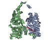 1zxnS S: Starting model for refinement C: citing same article ( |
|---|---|
| Similar structure data |


-Links
 PDBj
PDBj


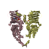
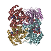
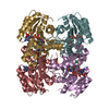
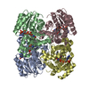
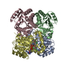
- Assembly
| Deposited unit | 
| ||||||||||||||||||||||||||||||||||||||||||||||||||||||||||||||||||||||||||||||||||||||||||||||||||||||||||||||||||||||||||||||||||||||||||||||||||||||
|---|---|---|---|---|---|---|---|---|---|---|---|---|---|---|---|---|---|---|---|---|---|---|---|---|---|---|---|---|---|---|---|---|---|---|---|---|---|---|---|---|---|---|---|---|---|---|---|---|---|---|---|---|---|---|---|---|---|---|---|---|---|---|---|---|---|---|---|---|---|---|---|---|---|---|---|---|---|---|---|---|---|---|---|---|---|---|---|---|---|---|---|---|---|---|---|---|---|---|---|---|---|---|---|---|---|---|---|---|---|---|---|---|---|---|---|---|---|---|---|---|---|---|---|---|---|---|---|---|---|---|---|---|---|---|---|---|---|---|---|---|---|---|---|---|---|---|---|---|---|---|---|
| 1 | 
| ||||||||||||||||||||||||||||||||||||||||||||||||||||||||||||||||||||||||||||||||||||||||||||||||||||||||||||||||||||||||||||||||||||||||||||||||||||||
| 2 | 
| ||||||||||||||||||||||||||||||||||||||||||||||||||||||||||||||||||||||||||||||||||||||||||||||||||||||||||||||||||||||||||||||||||||||||||||||||||||||
| Unit cell |
| ||||||||||||||||||||||||||||||||||||||||||||||||||||||||||||||||||||||||||||||||||||||||||||||||||||||||||||||||||||||||||||||||||||||||||||||||||||||
| Noncrystallographic symmetry (NCS) | NCS domain:
NCS domain segments: Component-ID: 0 / Beg auth comp-ID: SER / Beg label comp-ID: SER / Refine code: 0
NCS ensembles :
|
-Components
-Protein , 1 types, 4 molecules ABCD
| #1: Protein | Mass: 45660.535 Da / Num. of mol.: 4 / Fragment: UNP residues 29-428 Source method: isolated from a genetically manipulated source Source: (gene. exp.) Homo sapiens (human) / Gene: TOP2, TOP2A / Plasmid: pSB23 / Production host:  Escherichia coli (E. coli) / Strain (production host): Rosetta(DE3)pLysS / References: UniProt: P11388, EC: 5.99.1.3 Escherichia coli (E. coli) / Strain (production host): Rosetta(DE3)pLysS / References: UniProt: P11388, EC: 5.99.1.3 |
|---|
-Non-polymers , 5 types, 146 molecules 


| #2: Chemical | ChemComp-MG /  Mass: 24.305 Da / Num. of mol.: 4 / Source method: obtained synthetically / Formula: Mg Mass: 24.305 Da / Num. of mol.: 4 / Source method: obtained synthetically / Formula: Mg#3: Chemical | ChemComp-ADP / Adenosine diphosphate Mass: 427.201 Da / Num. of mol.: 4 / Source method: obtained synthetically / Formula: C10H15N5O10P2 / Comment: ADP, energy-carrying molecule*YM Mass: 427.201 Da / Num. of mol.: 4 / Source method: obtained synthetically / Formula: C10H15N5O10P2 / Comment: ADP, energy-carrying molecule*YM#4: Chemical | ChemComp-GOL / | Glycerol Mass: 92.094 Da / Num. of mol.: 1 / Source method: obtained synthetically / Formula: C3H8O3 Mass: 92.094 Da / Num. of mol.: 1 / Source method: obtained synthetically / Formula: C3H8O3#5: Chemical | Sulfate Mass: 96.063 Da / Num. of mol.: 3 / Source method: obtained synthetically / Formula: SO4 Mass: 96.063 Da / Num. of mol.: 3 / Source method: obtained synthetically / Formula: SO4#6: Water | ChemComp-HOH / | WaterMass: 18.015 Da / Num. of mol.: 134 / Source method: isolated from a natural source / Formula: H2O |
|---|
-Experimental details
-Experiment
| Experiment | Method: X-RAY DIFFRACTION / Number of used crystals: 1 |
|---|
- Sample preparation
| Crystal | Density Matthews: 2.61 Å3/Da / Density % sol: 52.85 % / Description: AUTHORS USED SF DATA FROM THE ENTRY 1ZXN. |
|---|
-Data collection
| Radiation | Protocol: SINGLE WAVELENGTH / Monochromatic (M) / Laue (L): M / Scattering type: x-ray |
|---|---|
| Radiation wavelength | Relative weight: 1 |
- Processing
| Software |
| |||||||||||||||||||||||||||||||||||||||||||||||||||||||||||||||||||||||||||
|---|---|---|---|---|---|---|---|---|---|---|---|---|---|---|---|---|---|---|---|---|---|---|---|---|---|---|---|---|---|---|---|---|---|---|---|---|---|---|---|---|---|---|---|---|---|---|---|---|---|---|---|---|---|---|---|---|---|---|---|---|---|---|---|---|---|---|---|---|---|---|---|---|---|---|---|---|
| Refinement | Method to determine structure: MOLECULAR REPLACEMENT Starting model: 1ZXN Resolution: 2.51→30 Å / Cor.coef. Fo:Fc: 0.945 / Cor.coef. Fo:Fc free: 0.929 / SU B: 10.968 / SU ML: 0.236 / Cross valid method: THROUGHOUT / σ(F): 0 / ESU R: 0.733 / ESU R Free: 0.299 / Stereochemistry target values: MAXIMUM LIKELIHOOD Details: HYDROGENS HAVE BEEN ADDED IN THE RIDING POSITIONS U VALUES : REFINED INDIVIDUALLY
| |||||||||||||||||||||||||||||||||||||||||||||||||||||||||||||||||||||||||||
| Solvent computation | Ion probe radii: 0.8 Å / Shrinkage radii: 0.8 Å / VDW probe radii: 1.2 Å / Solvent model: MASK | |||||||||||||||||||||||||||||||||||||||||||||||||||||||||||||||||||||||||||
| Displacement parameters | Biso max: 186.07 Å2 / Biso mean: 64.255 Å2 / Biso min: 32.38 Å2
| |||||||||||||||||||||||||||||||||||||||||||||||||||||||||||||||||||||||||||
| Refinement step | Cycle: LAST / Resolution: 2.51→30 Å
| |||||||||||||||||||||||||||||||||||||||||||||||||||||||||||||||||||||||||||
| Refine LS restraints |
| |||||||||||||||||||||||||||||||||||||||||||||||||||||||||||||||||||||||||||
| Refine LS restraints NCS | Refine-ID: X-RAY DIFFRACTION / Type: interatomic distance / Weight position: 0.05
| |||||||||||||||||||||||||||||||||||||||||||||||||||||||||||||||||||||||||||
| LS refinement shell | Resolution: 2.51→2.576 Å / Total num. of bins used: 20
|
