 Movie
Movie Controller

[English] 日本語
 Yorodumi
- PDB-4r0y: Structure of Maltose-binding Protein Fusion with the C-terminal G... -
+ Open data

- Basic information
| Entry | Database: PDB / ID: 4r0y | ||||||
|---|---|---|---|---|---|---|---|
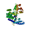
| Title | Structure of Maltose-binding Protein Fusion with the C-terminal GH1 domain of Guanylate Kinase-associated Protein from Rattus norvegicus | ||||||
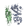
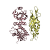
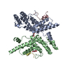
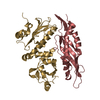
 Components Components | Maltose-binding periplasmic protein, Disks large-associated protein 1 | ||||||
 Keywords Keywords |  PROTEIN BINDING / three-helix bundle / synaptic scaffolding protein PROTEIN BINDING / three-helix bundle / synaptic scaffolding protein | ||||||
| Function / homology |  Function and homology information Function and homology informationmaintenance of postsynaptic density structure / postsynaptic specialization / Neurexins and neuroligins / signaling / structural constituent of postsynaptic density / protein localization to synapse / aggresome assembly / detection of maltose stimulus / maltose transport complex / maltose binding ...maintenance of postsynaptic density structure / postsynaptic specialization / Neurexins and neuroligins / signaling / structural constituent of postsynaptic density / protein localization to synapse / aggresome assembly / detection of maltose stimulus / maltose transport complex / maltose binding / maltose transport / maltodextrin transmembrane transport / carbohydrate transport / carbohydrate transmembrane transporter activity / ATP-binding cassette (ABC) transporter complex, substrate-binding subunit-containing / postsynaptic density, intracellular component / regulation of proteasomal protein catabolic process / ATP-binding cassette (ABC) transporter complex / cell chemotaxis / modulation of chemical synaptic transmission / outer membrane-bounded periplasmic space / postsynaptic membrane / postsynaptic density / periplasmic space / molecular adaptor activity / protein domain specific binding / glutamatergic synapse / synapse / DNA damage response / protein-containing complex binding / membrane / plasma membraneSimilarity search - Function | ||||||
| Biological species |  Escherichia coli K-12 (bacteria) Escherichia coli K-12 (bacteria) Rattus norvegicus (Norway rat) Rattus norvegicus (Norway rat) | ||||||
| Method | X-RAY DIFFRACTION / SYNCHROTRON / MOLECULAR REPLACEMENT / Resolution: 2 Å | ||||||
 Authors Authors | Im, Y.J. / Tong, J. | ||||||
 Citation Citation | Journal: Biochem.Biophys.Res.Commun. / Year: 2014 Title: Structure of the GH1 domain of guanylate kinase-associated protein from Rattus norvegicus. Authors: Tong, J. / Yang, H. / Eom, S.H. / Chun, C. / Im, Y.J. | ||||||
| History |
|
- Structure visualization
| Structure viewer | Molecule: MolmilJmol/JSmol |
|---|
- Downloads & links
-Download
| PDBx/mmCIF format | 4r0y.cif.gz | 195.2 KB | Display | PDBx/mmCIF format |
|---|---|---|---|---|
| PDB format | pdb4r0y.ent.gz | 155.1 KB | Display | PDB format |
| PDBx/mmJSON format | 4r0y.json.gz | Tree view | PDBx/mmJSON format | |
| Others |  Other downloads Other downloads |
-Validation report
| Arichive directory | https://data.pdbj.org/pub/pdb/validation_reports/r0/4r0yftp://data.pdbj.org/pub/pdb/validation_reports/r0/4r0y | HTTPS FTP |
|---|
-Related structure data
| Related structure data |  1ompS S: Starting model for refinement |
|---|---|
| Similar structure data |


-Links
 PDBj
PDBj


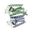
- Assembly
| Deposited unit | 
| ||||||||
|---|---|---|---|---|---|---|---|---|---|
| 1 | 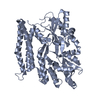
| ||||||||
| 2 | 
| ||||||||
| Unit cell |
|
-Components
| #1: Protein | Mass: 56217.723 Da / Num. of mol.: 2 Source method: isolated from a genetically manipulated source Details: Fusion protein of N-terminal residues 1-361 from Maltose-binding periplasmic protein (P0AEX9, MALE_ECOLI), linker residues (AM), residues 807-971 from Disks large-associated protein 1 ...Details: Fusion protein of N-terminal residues 1-361 from Maltose-binding periplasmic protein (P0AEX9, MALE_ECOLI), linker residues (AM), residues 807-971 from Disks large-associated protein 1 (P97836, DLGP1_RAT) with loop deletion of residue 917-945, the loop was replaced with residues VD. Source: (gene. exp.) Escherichia coli K-12 (bacteria), (gene. exp.) Rattus norvegicus (Norway rat)Plasmid: pHMBP / Production host: Escherichia coli (E. coli) / Strain (production host): BL21(DE3) / References: UniProt: P0AEX9, UniProt: P97836#2: Water | ChemComp-HOH / | Water Mass: 18.015 Da / Num. of mol.: 181 / Source method: isolated from a natural source / Formula: H2O Mass: 18.015 Da / Num. of mol.: 181 / Source method: isolated from a natural source / Formula: H2O |
|---|
-Experimental details
-Experiment
| Experiment | Method: X-RAY DIFFRACTION / Number of used crystals: 1 |
|---|
- Sample preparation
| Crystal | Density Matthews: 2.29 Å3/Da / Density % sol: 46.29 % |
|---|---|
| Crystal grow | Temperature: 298 K / Method: vapor diffusion, hanging drop / pH: 5 Details: 0.1M Sodium Citrate pH 5.0, 15% PEG 1500, 0.1M NaCl, VAPOR DIFFUSION, HANGING DROP, temperature 298K |
-Data collection
| Diffraction | Mean temperature: 100 K |
|---|---|
| Diffraction source | Source: SYNCHROTRON / Site: PAL/PLS  / Beamline: 7A (6B, 6C1) / Wavelength: 0.97857 Å / Beamline: 7A (6B, 6C1) / Wavelength: 0.97857 Å |
| Detector | Type: ADSC QUANTUM 270 / Detector: CCD / Date: Nov 2, 2013 / Details: mirrors |
| Radiation | Monochromator: Si 111 CHANNEL / Protocol: SINGLE WAVELENGTH / Monochromatic (M) / Laue (L): M / Scattering type: x-ray |
| Radiation wavelength | Wavelength: 0.97857 Å / Relative weight: 1 |
| Reflection | Resolution: 2→50 Å / Num. all: 72320 / Num. obs: 68103 / % possible obs: 97.1 % / Observed criterion σ(F): 3 / Observed criterion σ(I): 3 / Redundancy: 6.9 % / Biso Wilson estimate: 23.8 Å2 / Rmerge(I) obs: 0.076 / Net I/σ(I): 45.6 |
| Reflection shell | Resolution: 2→2.03 Å / Redundancy: 3.9 % / Rmerge(I) obs: 0.409 / Mean I/σ(I) obs: 3.92 / Num. unique all: 2793 / % possible all: 80.5 |
- Processing
| Software |
| ||||||||||||||||||||||||||||||||||||||||||||||||||||||||||||
|---|---|---|---|---|---|---|---|---|---|---|---|---|---|---|---|---|---|---|---|---|---|---|---|---|---|---|---|---|---|---|---|---|---|---|---|---|---|---|---|---|---|---|---|---|---|---|---|---|---|---|---|---|---|---|---|---|---|---|---|---|---|
| Refinement | Method to determine structure: MOLECULAR REPLACEMENT Starting model: PDB entry 1OMP Resolution: 2→39.52 Å / Rfactor Rfree error: 0.005 / Data cutoff high absF: 2315270.3 / Data cutoff low absF: 0 / Isotropic thermal model: RESTRAINED / Cross valid method: THROUGHOUT / σ(F): 0 / Stereochemistry target values: Engh & Huber
| ||||||||||||||||||||||||||||||||||||||||||||||||||||||||||||
| Solvent computation | Solvent model: FLAT MODEL / Bsol: 50.7122 Å2 / ksol: 0.367904 e/Å3 | ||||||||||||||||||||||||||||||||||||||||||||||||||||||||||||
| Displacement parameters | Biso mean: 48 Å2
| ||||||||||||||||||||||||||||||||||||||||||||||||||||||||||||
| Refine analyze |
| ||||||||||||||||||||||||||||||||||||||||||||||||||||||||||||
| Refinement step | Cycle: LAST / Resolution: 2→39.52 Å
| ||||||||||||||||||||||||||||||||||||||||||||||||||||||||||||
| Refine LS restraints |
| ||||||||||||||||||||||||||||||||||||||||||||||||||||||||||||
| LS refinement shell | Resolution: 2→2.13 Å / Rfactor Rfree error: 0.016 / Total num. of bins used: 6
| ||||||||||||||||||||||||||||||||||||||||||||||||||||||||||||
| Xplor file |
|
