 Movie
Movie Controller


+ Open data

- Basic information
| Entry | Database: PDB / ID: 4r0r | ||||||
|---|---|---|---|---|---|---|---|
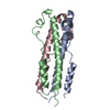
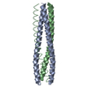
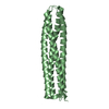
| Title | Ebolavirus GP Prehairpin Intermediate Mimic | ||||||
 Components Components | eboIZN21 | ||||||
 Keywords Keywords |  BIOSYNTHETIC PROTEIN / coiled-coil / N-trimer / prehairpin intermediate BIOSYNTHETIC PROTEIN / coiled-coil / N-trimer / prehairpin intermediate | ||||||
| Biological species | synthetic construct (others) | ||||||
| Method | X-RAY DIFFRACTION / SYNCHROTRON / MOLECULAR REPLACEMENT / molecular replacement / Resolution: 2.15 Å | ||||||
 Authors Authors | Clinton, T.R. / Weinstock, M.T. / Jacobsen, M.T. / Szabo-Fresnais, N. / Pandya, M.J. / Whitby, F.G. / Herbert, A.S. / Prugar, L.I. / McKinnon, R. / Hill, C.P. ...Clinton, T.R. / Weinstock, M.T. / Jacobsen, M.T. / Szabo-Fresnais, N. / Pandya, M.J. / Whitby, F.G. / Herbert, A.S. / Prugar, L.I. / McKinnon, R. / Hill, C.P. / Welch, B.D. / Dye, J.M. / Eckert, D.M. / Kay, M.S. | ||||||
 Citation Citation | Journal: Protein Sci. / Year: 2015 Title: Design and characterization of ebolavirus GP prehairpin intermediate mimics as drug targets. Authors: Clinton, T.R. / Weinstock, M.T. / Jacobsen, M.T. / Szabo-Fresnais, N. / Pandya, M.J. / Whitby, F.G. / Herbert, A.S. / Prugar, L.I. / McKinnon, R. / Hill, C.P. / Welch, B.D. / Dye, J.M. / Eckert, D.M. / Kay, M.S. | ||||||
| History |
|
- Structure visualization
| Structure viewer | Molecule:  MolmilJmol/JSmol MolmilJmol/JSmol |
|---|
- Downloads & links
-Download
| PDBx/mmCIF format | 4r0r.cif.gz | 29.1 KB | Display | PDBx/mmCIF format |
|---|---|---|---|---|
| PDB format | pdb4r0r.ent.gz | 20.5 KB | Display | PDB format |
| PDBx/mmJSON format | 4r0r.json.gz | Tree view | PDBx/mmJSON format | |
| Others |  Other downloads Other downloads |
-Validation report
| Arichive directory | https://data.pdbj.org/pub/pdb/validation_reports/r0/4r0rftp://data.pdbj.org/pub/pdb/validation_reports/r0/4r0r | HTTPS FTP |
|---|
-Related structure data
| Related structure data | |
|---|---|
| Similar structure data |


-Links
 PDBj
PDBj- Assembly
| Deposited unit | 
| ||||||||
|---|---|---|---|---|---|---|---|---|---|
| 1 | 
| ||||||||
| Unit cell |
| ||||||||
| Components on special symmetry positions |
|
-Components
| #1: Protein/peptide | Mass: 5621.546 Da / Num. of mol.: 1 / Source method: obtained synthetically / Source: (synth.) synthetic construct (others) |
|---|---|
| #2: Water | ChemComp-HOH / Water Mass: 18.015 Da / Num. of mol.: 6 / Source method: isolated from a natural source / Formula: H2O Mass: 18.015 Da / Num. of mol.: 6 / Source method: isolated from a natural source / Formula: H2O |
-Experimental details
-Experiment
| Experiment | Method: X-RAY DIFFRACTION / Number of used crystals: 1 |
|---|
- Sample preparation
| Crystal | Density Matthews: 2.76 Å3/Da / Density % sol: 55.5 % |
|---|---|
| Crystal grow | Temperature: 277 K / Method: vapor diffusion, sitting drop / pH: 7.5 Details: Synthetic peptide (protein) eboIZN21 dissolved in ddH2O at 10 mg/ml mixed in 2:1 protein:well buffer ratio with 30% (v/v) 1,2-propanediol, 100 mM HEPES pH 7.5, 20% (v/v) PEG-400 at 4 degrees ...Details: Synthetic peptide (protein) eboIZN21 dissolved in ddH2O at 10 mg/ml mixed in 2:1 protein:well buffer ratio with 30% (v/v) 1,2-propanediol, 100 mM HEPES pH 7.5, 20% (v/v) PEG-400 at 4 degrees Celcius (277 K), VAPOR DIFFUSION, SITTING DROP |
-Data collection
| Diffraction | Mean temperature: 100 K | |||||||||||||||||||||||||||||||||||||||||||||||||||||||||||||||||||||||||||||
|---|---|---|---|---|---|---|---|---|---|---|---|---|---|---|---|---|---|---|---|---|---|---|---|---|---|---|---|---|---|---|---|---|---|---|---|---|---|---|---|---|---|---|---|---|---|---|---|---|---|---|---|---|---|---|---|---|---|---|---|---|---|---|---|---|---|---|---|---|---|---|---|---|---|---|---|---|---|---|
| Diffraction source | Source: SYNCHROTRON / Site: SSRL  / Beamline: BL7-1 / Wavelength: 1.1 Å / Beamline: BL7-1 / Wavelength: 1.1 Å | |||||||||||||||||||||||||||||||||||||||||||||||||||||||||||||||||||||||||||||
| Detector | Type: ADSC QUANTUM 315r / Detector: CCD / Date: May 18, 2014 | |||||||||||||||||||||||||||||||||||||||||||||||||||||||||||||||||||||||||||||
| Radiation | Monochromator: Synchrotron / Protocol: SINGLE WAVELENGTH / Monochromatic (M) / Laue (L): M / Scattering type: x-ray | |||||||||||||||||||||||||||||||||||||||||||||||||||||||||||||||||||||||||||||
| Radiation wavelength | Wavelength: 1.1 Å / Relative weight: 1 | |||||||||||||||||||||||||||||||||||||||||||||||||||||||||||||||||||||||||||||
| Reflection | Resolution: 2.15→40 Å / Num. obs: 3680 / % possible obs: 99.9 % / Observed criterion σ(F): 0 / Observed criterion σ(I): 0 / Redundancy: 25.6 % / Biso Wilson estimate: 47.66 Å2 / Rmerge(I) obs: 0.054 / Χ2: 1.091 / Net I/σ(I): 16.7 | |||||||||||||||||||||||||||||||||||||||||||||||||||||||||||||||||||||||||||||
| Reflection shell |
|
-Phasing
| Phasing | Method: molecular replacement |
|---|
- Processing
| Software |
| ||||||||||||||||||||||||||||
|---|---|---|---|---|---|---|---|---|---|---|---|---|---|---|---|---|---|---|---|---|---|---|---|---|---|---|---|---|---|
| Refinement | Method to determine structure: MOLECULAR REPLACEMENT Starting model: CANONICAL HELICAL MODEL OF IZN AND N-TRIMER MODEL Resolution: 2.15→19.585 Å / SU ML: 0.37 / σ(F): 1.34 / Phase error: 42.49 / Stereochemistry target values: ML
| ||||||||||||||||||||||||||||
| Solvent computation | Shrinkage radii: 0.9 Å / VDW probe radii: 1.11 Å / Solvent model: FLAT BULK SOLVENT MODEL | ||||||||||||||||||||||||||||
| Displacement parameters | Biso max: 164.59 Å2 / Biso mean: 69.015 Å2 / Biso min: 40.63 Å2 | ||||||||||||||||||||||||||||
| Refinement step | Cycle: LAST / Resolution: 2.15→19.585 Å
| ||||||||||||||||||||||||||||
| Refine LS restraints |
| ||||||||||||||||||||||||||||
| LS refinement shell | Refine-ID: X-RAY DIFFRACTION / Total num. of bins used: 3
|
