 Movie
Movie Controller

[English] 日本語
 Yorodumi
- PDB-4qzu: Crystal Structure of wild type Human Cellular Retinol Binding Pro... -
+ Open data

- Basic information
| Entry | Database: PDB / ID: 4qzu | ||||||
|---|---|---|---|---|---|---|---|
| Title | Crystal Structure of wild type Human Cellular Retinol Binding Protein II (hCRBPII) bound to retinol at 11 KeV beam energy | ||||||
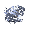
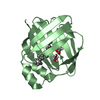
 Components Components | Retinol-binding protein 2 | ||||||
 Keywords Keywords |  PROTEIN BINDING / Retinol / Human Cellular Retinol Binding Protein II / intracellular lipid binding protein / X-ray flux / X-ray damage / Retinal / Retinoid Chaperones PROTEIN BINDING / Retinol / Human Cellular Retinol Binding Protein II / intracellular lipid binding protein / X-ray flux / X-ray damage / Retinal / Retinoid Chaperones | ||||||
| Function / homology |  Function and homology information Function and homology informationvitamin A metabolic process / retinoid binding / retinal binding / retinol binding / epidermis development / fatty acid transport / Retinoid metabolism and transport / fatty acid binding / nucleus / cytosolSimilarity search - Function | ||||||
| Biological species |  Homo sapiens (human) Homo sapiens (human) | ||||||
| Method | X-RAY DIFFRACTION / SYNCHROTRON / MOLECULAR REPLACEMENT / Resolution: 1.5 Å | ||||||
 Authors Authors | Assar, Z. / Geiger, J.H. | ||||||
 Citation Citation | Journal: Acta Crystallogr.,Sect.D / Year: 2014 Title: Structures of holo wild-type human cellular retinol-binding protein II (hCRBPII) bound to retinol and retinal. Authors: Nossoni, Z. / Assar, Z. / Yapici, I. / Nosrati, M. / Wang, W. / Berbasova, T. / Vasileiou, C. / Borhan, B. / Geiger, J. | ||||||
| History |
|
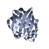
- Structure visualization
| Structure viewer | Molecule: MolmilJmol/JSmol |
|---|
- Downloads & links
-Download
| PDBx/mmCIF format | 4qzu.cif.gz | 249.3 KB | Display | PDBx/mmCIF format |
|---|---|---|---|---|
| PDB format | pdb4qzu.ent.gz | 202.2 KB | Display | PDB format |
| PDBx/mmJSON format | 4qzu.json.gz | Tree view | PDBx/mmJSON format | |
| Others |  Other downloads Other downloads |
-Validation report
| Arichive directory | https://data.pdbj.org/pub/pdb/validation_reports/qz/4qzuftp://data.pdbj.org/pub/pdb/validation_reports/qz/4qzu | HTTPS FTP |
|---|
-Related structure data
| Related structure data |  4qynC  4qypC  4qztC  2rctS S: Starting model for refinement C: citing same article ( |
|---|---|
| Similar structure data |


-Links
 PDBj
PDBj


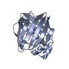
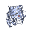
- Assembly
| Deposited unit | 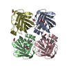
| ||||||||
|---|---|---|---|---|---|---|---|---|---|
| 1 | 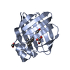
| ||||||||
| 2 | 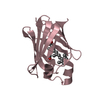
| ||||||||
| 3 | 
| ||||||||
| 4 | 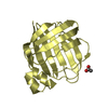
| ||||||||
| Unit cell |
|
-Components
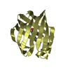
| #1: Protein | Mass: 15597.452 Da / Num. of mol.: 4 Source method: isolated from a genetically manipulated source Source: (gene. exp.) Homo sapiens (human) / Gene: RBP2, CRBP2 / Plasmid: Pet17b / Production host:  Escherichia coli (E. coli) / References: UniProt: P50120 Escherichia coli (E. coli) / References: UniProt: P50120#2: Chemical | Retinol  Mass: 286.452 Da / Num. of mol.: 3 / Source method: obtained synthetically / Formula: C20H30O Mass: 286.452 Da / Num. of mol.: 3 / Source method: obtained synthetically / Formula: C20H30O#3: Chemical | ChemComp-ACT / Acetate  Mass: 59.044 Da / Num. of mol.: 6 / Source method: obtained synthetically / Formula: C2H3O2 Mass: 59.044 Da / Num. of mol.: 6 / Source method: obtained synthetically / Formula: C2H3O2#4: Chemical | ChemComp-GOL / | Glycerol  Mass: 92.094 Da / Num. of mol.: 1 / Source method: obtained synthetically / Formula: C3H8O3 Mass: 92.094 Da / Num. of mol.: 1 / Source method: obtained synthetically / Formula: C3H8O3#5: Water | ChemComp-HOH / | Water Mass: 18.015 Da / Num. of mol.: 395 / Source method: isolated from a natural source / Formula: H2O Mass: 18.015 Da / Num. of mol.: 395 / Source method: isolated from a natural source / Formula: H2O |
|---|
-Experimental details
-Experiment
| Experiment | Method: X-RAY DIFFRACTION / Number of used crystals: 1 |
|---|
- Sample preparation
| Crystal | Density Matthews: 1.96 Å3/Da / Density % sol: 37.17 % |
|---|---|
| Crystal grow | Temperature: 298 K / pH: 4.5 Details: 25% PEG 4000 (40%), 0.1M Sodium Acetate pH 4.5, Ammonium Acetate 0.1M, VAPOR DIFFUSION, HANGING DROP, temperature 298K |
-Data collection
| Diffraction | Mean temperature: 100 K |
|---|---|
| Diffraction source | Source: SYNCHROTRON / Site: APS  / Beamline: 21-ID-D / Wavelength: 0.978 / Beamline: 21-ID-D / Wavelength: 0.978 |
| Detector | Type: MARMOSAIC 300 mm CCD / Detector: CCD / Date: Apr 20, 2014 |
| Radiation | Protocol: SINGLE WAVELENGTH / Monochromatic (M) / Laue (L): M / Scattering type: x-ray |
| Radiation wavelength | Wavelength: 0.978 Å / Relative weight: 1 |
| Reflection | Resolution: 1.496→34.66 Å / Num. obs: 76284 / % possible obs: 95.2 % / Redundancy: 4.9 % / Rmerge(I) obs: 0.042 / Net I/σ(I): 26.45 |
| Reflection shell | Resolution: 1.5→1.51 Å / Rmerge(I) obs: 0.598 / Mean I/σ(I) obs: 2.41 / % possible all: 88 |
- Processing
| Software |
| |||||||||||||||||||||||||||||||||||||||||||||||||||||||||||||||||||||||||||||||||||||||||||||||||||||||||||||||||||||||||||||||||||||||||||||||||||||||||||||||||||||||||||||||||||||||||||||
|---|---|---|---|---|---|---|---|---|---|---|---|---|---|---|---|---|---|---|---|---|---|---|---|---|---|---|---|---|---|---|---|---|---|---|---|---|---|---|---|---|---|---|---|---|---|---|---|---|---|---|---|---|---|---|---|---|---|---|---|---|---|---|---|---|---|---|---|---|---|---|---|---|---|---|---|---|---|---|---|---|---|---|---|---|---|---|---|---|---|---|---|---|---|---|---|---|---|---|---|---|---|---|---|---|---|---|---|---|---|---|---|---|---|---|---|---|---|---|---|---|---|---|---|---|---|---|---|---|---|---|---|---|---|---|---|---|---|---|---|---|---|---|---|---|---|---|---|---|---|---|---|---|---|---|---|---|---|---|---|---|---|---|---|---|---|---|---|---|---|---|---|---|---|---|---|---|---|---|---|---|---|---|---|---|---|---|---|---|---|---|
| Refinement | Method to determine structure: MOLECULAR REPLACEMENT Starting model: 2RCT Resolution: 1.5→34.66 Å / SU ML: 0.18 / σ(F): 1.97 / Phase error: 23.86 / Stereochemistry target values: ML
| |||||||||||||||||||||||||||||||||||||||||||||||||||||||||||||||||||||||||||||||||||||||||||||||||||||||||||||||||||||||||||||||||||||||||||||||||||||||||||||||||||||||||||||||||||||||||||||
| Solvent computation | Shrinkage radii: 0.9 Å / VDW probe radii: 1.11 Å / Solvent model: FLAT BULK SOLVENT MODEL | |||||||||||||||||||||||||||||||||||||||||||||||||||||||||||||||||||||||||||||||||||||||||||||||||||||||||||||||||||||||||||||||||||||||||||||||||||||||||||||||||||||||||||||||||||||||||||||
| Refinement step | Cycle: LAST / Resolution: 1.5→34.66 Å
| |||||||||||||||||||||||||||||||||||||||||||||||||||||||||||||||||||||||||||||||||||||||||||||||||||||||||||||||||||||||||||||||||||||||||||||||||||||||||||||||||||||||||||||||||||||||||||||
| Refine LS restraints |
| |||||||||||||||||||||||||||||||||||||||||||||||||||||||||||||||||||||||||||||||||||||||||||||||||||||||||||||||||||||||||||||||||||||||||||||||||||||||||||||||||||||||||||||||||||||||||||||
| LS refinement shell |
|
