 Movie
Movie Controller

[English] 日本語
 Yorodumi
- PDB-4qtc: Structure of human haspin (GSG2) in complex with SCH772984 reveal... -
+ Open data

- Basic information
| Entry | Database: PDB / ID: 4qtc | ||||||
|---|---|---|---|---|---|---|---|
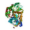
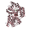
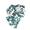
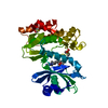
| Title | Structure of human haspin (GSG2) in complex with SCH772984 revealing the first type-I binding mode | ||||||
 Components Components | Serine/threonine-protein kinase haspin | ||||||
 Keywords Keywords | Transferase/transferase inhibitor /  Structural Genomics / Structural Genomics Consortium / SGC / TRANSFERASE / kinase / inhibitor / allosteric / Structural Genomics Consortium (SGC) / Transferase-transferase inhibitor complex Structural Genomics / Structural Genomics Consortium / SGC / TRANSFERASE / kinase / inhibitor / allosteric / Structural Genomics Consortium (SGC) / Transferase-transferase inhibitor complex | ||||||
| Function / homology |  Function and homology information Function and homology informationhistone H3T3 kinase activity / protein localization to chromosome, centromeric region / mitotic sister chromatid cohesion / mitotic spindle assembly checkpoint signaling / spindle / chromosome / mitotic cell cycle / non-specific serine/threonine protein kinase / protein kinase activity / intracellular signal transduction ...histone H3T3 kinase activity / protein localization to chromosome, centromeric region / mitotic sister chromatid cohesion / mitotic spindle assembly checkpoint signaling / spindle / chromosome / mitotic cell cycle / non-specific serine/threonine protein kinase / protein kinase activity / intracellular signal transduction / protein phosphorylation / protein serine kinase activity / centrosome / nucleoplasm / ATP binding / nucleus / cytoplasmSimilarity search - Function | ||||||
| Biological species |  Homo sapiens (human) Homo sapiens (human) | ||||||
| Method | X-RAY DIFFRACTION / SYNCHROTRON / MOLECULAR REPLACEMENT / Resolution: 1.4 Å | ||||||
 Authors Authors | Chaikuad, A. / von Delft, F. / Arrowsmith, C.H. / Edwards, A.M. / Bountra, C. / Knapp, S. / Structural Genomics Consortium (SGC) | ||||||
 Citation Citation | Journal: Nat.Chem.Biol. / Year: 2014 Title: A unique inhibitor binding site in ERK1/2 is associated with slow binding kinetics. Authors: Chaikuad, A. / M C Tacconi, E. / Zimmer, J. / Liang, Y. / Gray, N.S. / Tarsounas, M. / Knapp, S. | ||||||
| History |
|
- Structure visualization
| Structure viewer | Molecule: MolmilJmol/JSmol |
|---|
- Downloads & links
-Download
| PDBx/mmCIF format | 4qtc.cif.gz | 163.1 KB | Display | PDBx/mmCIF format |
|---|---|---|---|---|
| PDB format | pdb4qtc.ent.gz | 127.1 KB | Display | PDB format |
| PDBx/mmJSON format | 4qtc.json.gz | Tree view | PDBx/mmJSON format | |
| Others |  Other downloads Other downloads |
-Validation report
| Arichive directory | https://data.pdbj.org/pub/pdb/validation_reports/qt/4qtcftp://data.pdbj.org/pub/pdb/validation_reports/qt/4qtc | HTTPS FTP |
|---|
-Related structure data
| Related structure data |  4qtaC  4qtbC  4qtdC  4qteC  3dlzS C: citing same article ( S: Starting model for refinement |
|---|---|
| Similar structure data |


-Links
 PDBj
PDBj- Assembly
| Deposited unit | 
| ||||||||
|---|---|---|---|---|---|---|---|---|---|
| 1 |
| ||||||||
| Unit cell |
|
-Components
| #1: Protein | Mass: 40711.484 Da / Num. of mol.: 1 / Fragment: kinase domain (465-798) Source method: isolated from a genetically manipulated source Source: (gene. exp.) Homo sapiens (human) / Gene: GSG2 / Plasmid: pNIC28-Bsa4 / Production host:  Escherichia coli (E. coli) / Strain (production host): BL21-R3-pRARE2 Escherichia coli (E. coli) / Strain (production host): BL21-R3-pRARE2References: UniProt: Q8TF76, non-specific serine/threonine protein kinase | ||||
|---|---|---|---|---|---|
| #2: Chemical | ChemComp-MPD / (2-Methyl-2,4-pentanediol  Mass: 118.174 Da / Num. of mol.: 1 / Source method: obtained synthetically / Formula: C6H14O2 / Comment: precipitant*YM Mass: 118.174 Da / Num. of mol.: 1 / Source method: obtained synthetically / Formula: C6H14O2 / Comment: precipitant*YM | ||||
| #3: Chemical | Glycerol  Mass: 92.094 Da / Num. of mol.: 2 / Source method: obtained synthetically / Formula: C3H8O3 Mass: 92.094 Da / Num. of mol.: 2 / Source method: obtained synthetically / Formula: C3H8O3#4: Chemical | ChemComp-38Z / ( |   Mass: 587.674 Da / Num. of mol.: 1 / Source method: obtained synthetically / Formula: C33H33N9O2 Mass: 587.674 Da / Num. of mol.: 1 / Source method: obtained synthetically / Formula: C33H33N9O2#5: Water | ChemComp-HOH / | Water Mass: 18.015 Da / Num. of mol.: 500 / Source method: isolated from a natural source / Formula: H2O Mass: 18.015 Da / Num. of mol.: 500 / Source method: isolated from a natural source / Formula: H2O |
-Experimental details
-Experiment
| Experiment | Method: X-RAY DIFFRACTION / Number of used crystals: 1 |
|---|
- Sample preparation
| Crystal | Density Matthews: 3.06 Å3/Da / Density % sol: 59.86 % |
|---|---|
| Crystal grow | Temperature: 277.15 K / Method: vapor diffusion, sitting drop / pH: 6 Details: 51% MPD and 0.1 M SPG pH 6.0, VAPOR DIFFUSION, SITTING DROP, temperature 277.15K |
-Data collection
| Diffraction | Mean temperature: 100 K |
|---|---|
| Diffraction source | Source: SYNCHROTRON / Site: Diamond  / Beamline: I02 / Wavelength: 0.97949 Å / Beamline: I02 / Wavelength: 0.97949 Å |
| Detector | Type: PSI PILATUS 6M / Detector: PIXEL / Date: Oct 24, 2013 / Details: Kirkpatrick Baez bimorph mirror pair |
| Radiation | Monochromator: Si (111) double crystal monochromator / Protocol: SINGLE WAVELENGTH / Monochromatic (M) / Laue (L): M / Scattering type: x-ray |
| Radiation wavelength | Wavelength: 0.97949 Å / Relative weight: 1 |
| Reflection | Resolution: 1.4→36.24 Å / Num. all: 99002 / Num. obs: 98923 / % possible obs: 100 % / Observed criterion σ(F): 0 / Observed criterion σ(I): 0 / Redundancy: 6.1 % / Biso Wilson estimate: 17.4 Å2 / Rmerge(I) obs: 0.044 / Net I/σ(I): 17.9 |
| Reflection shell | Resolution: 1.4→1.48 Å / Redundancy: 6.3 % / Rmerge(I) obs: 0.693 / Mean I/σ(I) obs: 2.4 / Num. unique all: 14277 / % possible all: 100 |
- Processing
| Software |
| |||||||||||||||||||||||||||||||||||||||||||||||||||||||||||||||||||||||||||||||||||||||||||||||||||||||||
|---|---|---|---|---|---|---|---|---|---|---|---|---|---|---|---|---|---|---|---|---|---|---|---|---|---|---|---|---|---|---|---|---|---|---|---|---|---|---|---|---|---|---|---|---|---|---|---|---|---|---|---|---|---|---|---|---|---|---|---|---|---|---|---|---|---|---|---|---|---|---|---|---|---|---|---|---|---|---|---|---|---|---|---|---|---|---|---|---|---|---|---|---|---|---|---|---|---|---|---|---|---|---|---|---|---|---|
| Refinement | Method to determine structure: MOLECULAR REPLACEMENT Starting model: pdb entry 3DLZ Resolution: 1.4→36.19 Å / Cor.coef. Fo:Fc: 0.979 / Cor.coef. Fo:Fc free: 0.975 / SU B: 1.567 / SU ML: 0.031 / Cross valid method: THROUGHOUT / σ(F): 0 / σ(I): 2 / ESU R: 0.043 / ESU R Free: 0.044 / Stereochemistry target values: MAXIMUM LIKELIHOOD / Details: HYDROGENS HAVE BEEN ADDED IN THE RIDING POSITIONS
| |||||||||||||||||||||||||||||||||||||||||||||||||||||||||||||||||||||||||||||||||||||||||||||||||||||||||
| Solvent computation | Ion probe radii: 0.8 Å / Shrinkage radii: 0.8 Å / VDW probe radii: 1.2 Å / Solvent model: MASK | |||||||||||||||||||||||||||||||||||||||||||||||||||||||||||||||||||||||||||||||||||||||||||||||||||||||||
| Displacement parameters | Biso mean: 26.339 Å2
| |||||||||||||||||||||||||||||||||||||||||||||||||||||||||||||||||||||||||||||||||||||||||||||||||||||||||
| Refine analyze | Luzzati coordinate error obs: 0.16 Å | |||||||||||||||||||||||||||||||||||||||||||||||||||||||||||||||||||||||||||||||||||||||||||||||||||||||||
| Refinement step | Cycle: LAST / Resolution: 1.4→36.19 Å
| |||||||||||||||||||||||||||||||||||||||||||||||||||||||||||||||||||||||||||||||||||||||||||||||||||||||||
| Refine LS restraints |
| |||||||||||||||||||||||||||||||||||||||||||||||||||||||||||||||||||||||||||||||||||||||||||||||||||||||||
| LS refinement shell | Resolution: 1.4→1.436 Å / Total num. of bins used: 20
| |||||||||||||||||||||||||||||||||||||||||||||||||||||||||||||||||||||||||||||||||||||||||||||||||||||||||
| Refinement TLS params. | Method: refined / Refine-ID: X-RAY DIFFRACTION
| |||||||||||||||||||||||||||||||||||||||||||||||||||||||||||||||||||||||||||||||||||||||||||||||||||||||||
| Refinement TLS group |
|
