| Entry | Database: PDB / ID: 4qpx
|
|---|
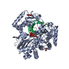
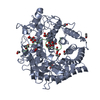
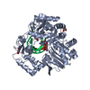
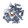
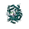
| Title | NV polymerase post-incorporation-like complex |
|---|
 Components Components | - Polyprotein
 Proteolysis Proteolysis - RNA (5'-R(*U*AP*CP*CP*CP*GP*GP*G)-3')
- RNA (5'-R(*UP*GP*CP*CP*CP*GP*GP*G)-3')
|
|---|
 Keywords Keywords | Hydrolase/RNA / RNA-depedent RNA polymerase / Hydrolase-RNA complex |
|---|
| Function / homology |  Function and homology information Function and homology information |
|---|
| Biological species |  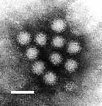 Norwalk virus Norwalk virus |
|---|
| Method | X-RAY DIFFRACTION / SYNCHROTRON / MOLECULAR REPLACEMENT / Resolution: 1.86 Å |
|---|
 Authors Authors | Zamyatkin, D.F. / Parra, F. / Grochulski, P. / Ng, K.K.S. |
|---|
 Citation Citation | Journal: Acta Crystallogr.,Sect.D / Year: 2014
Title: Structure of a backtracked state reveals conformational changes similar to the state following nucleotide incorporation in human norovirus polymerase.
Authors: Zamyatkin, D. / Rao, C. / Hoffarth, E. / Jurca, G. / Rho, H. / Parra, F. / Grochulski, P. / Ng, K.K. |
|---|
| History | | Deposition | Jun 25, 2014 | Deposition site: RCSB / Processing site: RCSB |
|---|
| Revision 1.0 | Apr 22, 2015 | Provider: repository / Type: Initial release |
|---|
| Revision 1.1 | Sep 20, 2023 | Group: Data collection / Database references ...Data collection / Database references / Derived calculations / Refinement description
Category: chem_comp_atom / chem_comp_bond ...chem_comp_atom / chem_comp_bond / database_2 / pdbx_initial_refinement_model / pdbx_struct_conn_angle / struct_conn / struct_ref_seq_dif / struct_site
Item: _database_2.pdbx_DOI / _database_2.pdbx_database_accession ..._database_2.pdbx_DOI / _database_2.pdbx_database_accession / _pdbx_struct_conn_angle.ptnr1_auth_asym_id / _pdbx_struct_conn_angle.ptnr1_auth_comp_id / _pdbx_struct_conn_angle.ptnr1_auth_seq_id / _pdbx_struct_conn_angle.ptnr1_label_asym_id / _pdbx_struct_conn_angle.ptnr1_label_atom_id / _pdbx_struct_conn_angle.ptnr1_label_comp_id / _pdbx_struct_conn_angle.ptnr1_label_seq_id / _pdbx_struct_conn_angle.ptnr3_auth_asym_id / _pdbx_struct_conn_angle.ptnr3_auth_comp_id / _pdbx_struct_conn_angle.ptnr3_auth_seq_id / _pdbx_struct_conn_angle.ptnr3_label_asym_id / _pdbx_struct_conn_angle.ptnr3_label_atom_id / _pdbx_struct_conn_angle.ptnr3_label_comp_id / _pdbx_struct_conn_angle.ptnr3_label_seq_id / _pdbx_struct_conn_angle.value / _struct_conn.pdbx_dist_value / _struct_conn.ptnr1_auth_asym_id / _struct_conn.ptnr1_auth_comp_id / _struct_conn.ptnr1_auth_seq_id / _struct_conn.ptnr1_label_asym_id / _struct_conn.ptnr1_label_atom_id / _struct_conn.ptnr1_label_comp_id / _struct_conn.ptnr1_label_seq_id / _struct_conn.ptnr2_auth_asym_id / _struct_conn.ptnr2_auth_comp_id / _struct_conn.ptnr2_auth_seq_id / _struct_conn.ptnr2_label_asym_id / _struct_conn.ptnr2_label_atom_id / _struct_conn.ptnr2_label_comp_id / _struct_conn.ptnr2_label_seq_id / _struct_ref_seq_dif.details / _struct_site.pdbx_auth_asym_id / _struct_site.pdbx_auth_comp_id / _struct_site.pdbx_auth_seq_id |
|---|
|
|---|
 Movie
Movie Controller


 Open data
 Basic information
 Structure visualization
 Downloads & links
 Other downloads


 PDBj
PDBj Assembly
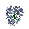


 Mass: 92.094 Da / Num. of mol.: 2 / Source method: obtained synthetically / Formula: C3H8O3
 Mass: 54.938 Da / Num. of mol.: 3 / Source method: obtained synthetically / Formula: Mn
 Sample preparation
 / Beamline: 08ID-1 / Wavelength: 0.98 Å
 Processing