 Movie
Movie Controller

[English] 日本語
 Yorodumi
- PDB-4qo0: Crystal structure of rhomboid intramembrane protease GlpG in comp... -
+ Open data

- Basic information
| Entry | Database: PDB / ID: 4qo0 | ||||||
|---|---|---|---|---|---|---|---|
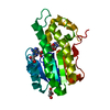
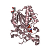
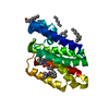
| Title | Crystal structure of rhomboid intramembrane protease GlpG in complex with peptide derived inhibitor Ac-FATA-cmk | ||||||
 Components Components |
| ||||||
 Keywords Keywords | HYDROLASE/HYDROLASE INHIBITOR /  alpha-helical / Rhomboid intramembrane protease / HYDROLASE-HYDROLASE INHIBITOR complex alpha-helical / Rhomboid intramembrane protease / HYDROLASE-HYDROLASE INHIBITOR complex | ||||||
| Function / homology | Rhomboid-like fold / Rhomboid-like / Up-down Bundle / Mainly Alpha / ACE-PHE-ALA-THR-ALA-0QE / :  Function and homology information Function and homology information | ||||||
| Biological species |  Escherichia coli (E. coli) Escherichia coli (E. coli) | ||||||
| Method | X-RAY DIFFRACTION / SYNCHROTRON / MOLECULAR REPLACEMENT / Resolution: 2.9 Å | ||||||
 Authors Authors | Zoll, S. / Strisovsky, K. | ||||||
 Citation Citation | Journal: Embo J. / Year: 2014 Title: Substrate binding and specificity of rhomboid intramembrane protease revealed by substrate-peptide complex structures. Authors: Zoll, S. / Stanchev, S. / Began, J. / Skerle, J. / Lepsik, M. / Peclinovska, L. / Majer, P. / Strisovsky, K. | ||||||
| History |
|
- Structure visualization
| Structure viewer | Molecule: MolmilJmol/JSmol |
|---|
- Downloads & links
-Download
| PDBx/mmCIF format | 4qo0.cif.gz | 53.8 KB | Display | PDBx/mmCIF format |
|---|---|---|---|---|
| PDB format | pdb4qo0.ent.gz | 36.9 KB | Display | PDB format |
| PDBx/mmJSON format | 4qo0.json.gz | Tree view | PDBx/mmJSON format | |
| Others |  Other downloads Other downloads |
-Validation report
| Arichive directory | https://data.pdbj.org/pub/pdb/validation_reports/qo/4qo0ftp://data.pdbj.org/pub/pdb/validation_reports/qo/4qo0 | HTTPS FTP |
|---|
-Related structure data
| Related structure data |  4qnzC  4qo2C 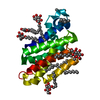 2ic8S C: citing same article ( S: Starting model for refinement |
|---|---|
| Similar structure data |


-Links
 PDBj
PDBj- Assembly
| Deposited unit | 
| ||||||||
|---|---|---|---|---|---|---|---|---|---|
| 1 |
| ||||||||
| 2 | 
| ||||||||
| Unit cell |
|
-Components
| #1: Protein | Mass: 22737.914 Da / Num. of mol.: 1 / Fragment: Rhomboid protease GlpG Source method: isolated from a genetically manipulated source Source: (gene. exp.) Escherichia coli (E. coli) / Strain: C41 / Gene: glpG, BN896_3117 / Production host: Escherichia coli (E. coli) / References: UniProt: U6NA71, rhomboid protease | ||
|---|---|---|---|
| #2: Protein/peptide |   Type: Peptide-like / Class: Inhibitor / Mass: 466.959 Da / Num. of mol.: 1 / Source method: obtained synthetically / References: ACE-PHE-ALA-THR-ALA-0QE Type: Peptide-like / Class: Inhibitor / Mass: 466.959 Da / Num. of mol.: 1 / Source method: obtained synthetically / References: ACE-PHE-ALA-THR-ALA-0QE | ||
| #3: Chemical | ChemComp-CL / Chloride  Mass: 35.453 Da / Num. of mol.: 1 / Source method: obtained synthetically / Formula: Cl Mass: 35.453 Da / Num. of mol.: 1 / Source method: obtained synthetically / Formula: Cl | ||
| #4: Sugar |   Type: D-saccharide / Mass: 306.395 Da / Num. of mol.: 2 Type: D-saccharide / Mass: 306.395 Da / Num. of mol.: 2Source method: isolated from a genetically manipulated source Formula: C15H30O6 / Comment: detergent*YM #5: Water | ChemComp-HOH / | Water Mass: 18.015 Da / Num. of mol.: 5 / Source method: isolated from a natural source / Formula: H2O Mass: 18.015 Da / Num. of mol.: 5 / Source method: isolated from a natural source / Formula: H2O |
-Experimental details
-Experiment
| Experiment | Method: X-RAY DIFFRACTION / Number of used crystals: 1 |
|---|
- Sample preparation
| Crystal | Density Matthews: 3.93 Å3/Da / Density % sol: 68.66 % |
|---|---|
| Crystal grow | Temperature: 293 K / Method: vapor diffusion, sitting drop / pH: 9.5 Details: 10% PEG 8000, 0.1 M CHES pH 9.5, 0.2 M sodium chloride, VAPOR DIFFUSION, SITTING DROP, temperature 293K |
-Data collection
| Diffraction | Mean temperature: 100 K |
|---|---|
| Diffraction source | Source: SYNCHROTRON / Site: ESRF  / Beamline: ID29 / Wavelength: 1 Å / Beamline: ID29 / Wavelength: 1 Å |
| Detector | Type: DECTRIS PILATUS 6M-F / Detector: PIXEL / Date: Oct 3, 2013 |
| Radiation | Monochromator: Si 111 CHANNEL / Protocol: SINGLE WAVELENGTH / Monochromatic (M) / Laue (L): M / Scattering type: x-ray |
| Radiation wavelength | Wavelength: 1 Å / Relative weight: 1 |
| Reflection | Resolution: 2.9→51.79 Å / Num. all: 8094 / Num. obs: 8086 / % possible obs: 99.9 % / Observed criterion σ(F): -3 / Observed criterion σ(I): -3 |
| Reflection shell | Resolution: 2.9→3.003 Å / % possible all: 100 |
- Processing
| Software |
| ||||||||||||||||||||||||||||||||||||||||||||||||||||||||||||||||||||||||||||||||||||||||||||||||||||||||||||||||||||||||||||||||||||||||||||||||||||||||||||||||||||||||||||||||||||||
|---|---|---|---|---|---|---|---|---|---|---|---|---|---|---|---|---|---|---|---|---|---|---|---|---|---|---|---|---|---|---|---|---|---|---|---|---|---|---|---|---|---|---|---|---|---|---|---|---|---|---|---|---|---|---|---|---|---|---|---|---|---|---|---|---|---|---|---|---|---|---|---|---|---|---|---|---|---|---|---|---|---|---|---|---|---|---|---|---|---|---|---|---|---|---|---|---|---|---|---|---|---|---|---|---|---|---|---|---|---|---|---|---|---|---|---|---|---|---|---|---|---|---|---|---|---|---|---|---|---|---|---|---|---|---|---|---|---|---|---|---|---|---|---|---|---|---|---|---|---|---|---|---|---|---|---|---|---|---|---|---|---|---|---|---|---|---|---|---|---|---|---|---|---|---|---|---|---|---|---|---|---|---|---|
| Refinement | Method to determine structure: MOLECULAR REPLACEMENT Starting model: PDB ENTRY 2IC8 Resolution: 2.9→51.79 Å / Cor.coef. Fo:Fc: 0.945 / Cor.coef. Fo:Fc free: 0.921 / SU B: 10.636 / SU ML: 0.202 / Cross valid method: THROUGHOUT / ESU R: 0.534 / ESU R Free: 0.296 / Stereochemistry target values: MAXIMUM LIKELIHOOD / Details: HYDROGENS HAVE BEEN ADDED IN THE RIDING POSITIONS
| ||||||||||||||||||||||||||||||||||||||||||||||||||||||||||||||||||||||||||||||||||||||||||||||||||||||||||||||||||||||||||||||||||||||||||||||||||||||||||||||||||||||||||||||||||||||
| Solvent computation | Ion probe radii: 0.7 Å / Shrinkage radii: 0.7 Å / VDW probe radii: 1.2 Å / Solvent model: MASK | ||||||||||||||||||||||||||||||||||||||||||||||||||||||||||||||||||||||||||||||||||||||||||||||||||||||||||||||||||||||||||||||||||||||||||||||||||||||||||||||||||||||||||||||||||||||
| Displacement parameters | Biso mean: 54.105 Å2
| ||||||||||||||||||||||||||||||||||||||||||||||||||||||||||||||||||||||||||||||||||||||||||||||||||||||||||||||||||||||||||||||||||||||||||||||||||||||||||||||||||||||||||||||||||||||
| Refinement step | Cycle: LAST / Resolution: 2.9→51.79 Å
| ||||||||||||||||||||||||||||||||||||||||||||||||||||||||||||||||||||||||||||||||||||||||||||||||||||||||||||||||||||||||||||||||||||||||||||||||||||||||||||||||||||||||||||||||||||||
| Refine LS restraints |
|