 Movie
Movie Controller

[English] 日本語
 Yorodumi
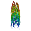
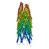
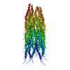
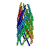
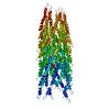
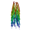
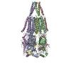
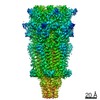
- PDB-4qnl: Crystal structure of tail fiber protein gp63.1 from E. coli phage G7C -
+ Open data

- Basic information
| Entry | Database: PDB / ID: 4qnl | ||||||
|---|---|---|---|---|---|---|---|
| Title | Crystal structure of tail fiber protein gp63.1 from E. coli phage G7C | ||||||
 Components Components | Tail fiber protein | ||||||
 Keywords Keywords |  HYDROLASE / tail fiber / G7C phage / hydrolase-type esterase / SGNH hydrolase-type esterase domain (IPR013831) / Adsorption of the phage on bacterial host / bacterial LPS digestion / tail fiber protein gp66 / selenomethionine derivative / distal end of the baseplate HYDROLASE / tail fiber / G7C phage / hydrolase-type esterase / SGNH hydrolase-type esterase domain (IPR013831) / Adsorption of the phage on bacterial host / bacterial LPS digestion / tail fiber protein gp66 / selenomethionine derivative / distal end of the baseplate | ||||||
| Function / homology |  Function and homology informationSeminal Fluid Protein PDC-109 (Domain B) - #80 / NE0471 N-terminal domain-like - #50 / NE0471 N-terminal domain-like / Tail spike TSP1/Gp66, N-terminal domain / Tail spike TSP1/Gp66 receptor binding N-terminal domain / Seminal Fluid Protein PDC-109 (Domain B) / Ribbon / 2-Layer Sandwich / Mainly Beta / Alpha Beta Function and homology informationSeminal Fluid Protein PDC-109 (Domain B) - #80 / NE0471 N-terminal domain-like - #50 / NE0471 N-terminal domain-like / Tail spike TSP1/Gp66, N-terminal domain / Tail spike TSP1/Gp66 receptor binding N-terminal domain / Seminal Fluid Protein PDC-109 (Domain B) / Ribbon / 2-Layer Sandwich / Mainly Beta / Alpha BetaSimilarity search - Domain/homology | ||||||
| Biological species |  Escherichia phage vB_EcoP_G7C (virus) Escherichia phage vB_EcoP_G7C (virus) | ||||||
| Method | X-RAY DIFFRACTION / SYNCHROTRON / SAD / Resolution: 2.411 Å | ||||||
 Authors Authors | Riccio, C. / Browning, C. / Prokhorov, N. / Letarov, A. / Leiman, P.G. | ||||||
 Citation Citation | Journal: Mol. Microbiol. / Year: 2017 Title: Function of bacteriophage G7C esterase tailspike in host cell adsorption. Authors: Prokhorov, N.S. / Riccio, C. / Zdorovenko, E.L. / Shneider, M.M. / Browning, C. / Knirel, Y.A. / Leiman, P.G. / Letarov, A.V. | ||||||
| History |
|
- Structure visualization
| Structure viewer | Molecule: MolmilJmol/JSmol |
|---|
- Downloads & links
-Download
| PDBx/mmCIF format | 4qnl.cif.gz | 337.8 KB | Display | PDBx/mmCIF format |
|---|---|---|---|---|
| PDB format | pdb4qnl.ent.gz | 286.7 KB | Display | PDB format |
| PDBx/mmJSON format | 4qnl.json.gz | Tree view | PDBx/mmJSON format | |
| Others |  Other downloads Other downloads |
-Validation report
| Arichive directory | https://data.pdbj.org/pub/pdb/validation_reports/qn/4qnlftp://data.pdbj.org/pub/pdb/validation_reports/qn/4qnl | HTTPS FTP |
|---|
-Related structure data
| Similar structure data |
|---|
-Links
 PDBj
PDBj- Assembly
| Deposited unit | 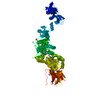
| |||||||||||||||||||||
|---|---|---|---|---|---|---|---|---|---|---|---|---|---|---|---|---|---|---|---|---|---|---|
| 1 | 
| |||||||||||||||||||||
| Unit cell |
| |||||||||||||||||||||
| Components on special symmetry positions |
|
-Components
-Protein , 1 types, 1 molecules A
| #1: Protein | Mass: 94838.531 Da / Num. of mol.: 1 Source method: isolated from a genetically manipulated source Source: (gene. exp.) Escherichia phage vB_EcoP_G7C (virus) / Gene: 63.1, gp63.1 / Plasmid: pET-23a / Production host:  Escherichia coli (E. coli) / Strain (production host): B834 / References: UniProt: G0XNW5 Escherichia coli (E. coli) / Strain (production host): B834 / References: UniProt: G0XNW5 |
|---|
-Non-polymers , 6 types, 341 molecules 


| #2: Chemical | Diethylene glycol Mass: 106.120 Da / Num. of mol.: 3 / Source method: obtained synthetically / Formula: C4H10O3 Mass: 106.120 Da / Num. of mol.: 3 / Source method: obtained synthetically / Formula: C4H10O3#3: Chemical | Ethylene glycol Mass: 62.068 Da / Num. of mol.: 2 / Source method: obtained synthetically / Formula: C2H6O2 Mass: 62.068 Da / Num. of mol.: 2 / Source method: obtained synthetically / Formula: C2H6O2#4: Chemical | ChemComp-ZN / |  Mass: 65.409 Da / Num. of mol.: 1 / Source method: obtained synthetically / Formula: Zn Mass: 65.409 Da / Num. of mol.: 1 / Source method: obtained synthetically / Formula: Zn#5: Chemical | ChemComp-SO4 / | Sulfate Mass: 96.063 Da / Num. of mol.: 1 / Source method: obtained synthetically / Formula: SO4 Mass: 96.063 Da / Num. of mol.: 1 / Source method: obtained synthetically / Formula: SO4#6: Chemical | ChemComp-CL / Chloride Mass: 35.453 Da / Num. of mol.: 4 / Source method: obtained synthetically / Formula: Cl Mass: 35.453 Da / Num. of mol.: 4 / Source method: obtained synthetically / Formula: Cl#7: Water | ChemComp-HOH / | WaterMass: 18.015 Da / Num. of mol.: 330 / Source method: isolated from a natural source / Formula: H2O |
|---|
-Experimental details
-Experiment
| Experiment | Method: X-RAY DIFFRACTION / Number of used crystals: 1 |
|---|
- Sample preparation
| Crystal | Density Matthews: 2.41 Å3/Da / Density % sol: 48.96 % |
|---|---|
| Crystal grow | Temperature: 293 K / Method: vapor diffusion, hanging drop / pH: 5.5 Details: 150 mM Ammonium Sulphate, 34% PEE 797, 100 mM Bis-Tris pH 5.5; 1.5 ul protein @ 14.3 mg/ml + 1.5 ul well solution, VAPOR DIFFUSION, HANGING DROP, temperature 293K |
-Data collection
| Diffraction | Mean temperature: 100 K |
|---|---|
| Diffraction source | Source: SYNCHROTRON / Site: SLS  / Beamline: X06SA / Wavelength: 0.97969 Å / Beamline: X06SA / Wavelength: 0.97969 Å |
| Detector | Type: DECTRIS PILATUS 6M / Detector: PIXEL / Date: Nov 4, 2012 / Details: dynamically bendable mirror |
| Radiation | Monochromator: Si(111) monochromator / Protocol: SINGLE WAVELENGTH / Monochromatic (M) / Laue (L): M / Scattering type: x-ray |
| Radiation wavelength | Wavelength: 0.97969 Å / Relative weight: 1 |
| Reflection | Resolution: 2.4→92 Å / Num. all: 67757 / Num. obs: 67757 / % possible obs: 99.1 % / Observed criterion σ(F): 0 / Observed criterion σ(I): 2 / Redundancy: 5.1 % / Biso Wilson estimate: 57.1 Å2 / Rmerge(I) obs: 0.075 / Net I/σ(I): 15.9 |
| Reflection shell | Resolution: 2.41→2.56 Å / Redundancy: 4.4 % / Rmerge(I) obs: 0.72 / Mean I/σ(I) obs: 2 / Num. unique all: 10495 / % possible all: 94.4 |
- Processing
| Software |
| ||||||||||||||||||||||||||||||||||||||||||||||||||||||||||||||||||||||||||||||||||||||||||||||||||||||||||||||||||||||||||||||||||||||||||||||||||||||||||||||||||||||||||||||||||||||||||||||||||||||||
|---|---|---|---|---|---|---|---|---|---|---|---|---|---|---|---|---|---|---|---|---|---|---|---|---|---|---|---|---|---|---|---|---|---|---|---|---|---|---|---|---|---|---|---|---|---|---|---|---|---|---|---|---|---|---|---|---|---|---|---|---|---|---|---|---|---|---|---|---|---|---|---|---|---|---|---|---|---|---|---|---|---|---|---|---|---|---|---|---|---|---|---|---|---|---|---|---|---|---|---|---|---|---|---|---|---|---|---|---|---|---|---|---|---|---|---|---|---|---|---|---|---|---|---|---|---|---|---|---|---|---|---|---|---|---|---|---|---|---|---|---|---|---|---|---|---|---|---|---|---|---|---|---|---|---|---|---|---|---|---|---|---|---|---|---|---|---|---|---|---|---|---|---|---|---|---|---|---|---|---|---|---|---|---|---|---|---|---|---|---|---|---|---|---|---|---|---|---|---|---|---|---|
| Refinement | Method to determine structure: SAD / Resolution: 2.411→64.959 Å / SU ML: 0.29 / Isotropic thermal model: Isotropic with TLS / Cross valid method: FREE R / σ(F): 1.21 / Phase error: 23.28 / Stereochemistry target values: ML
| ||||||||||||||||||||||||||||||||||||||||||||||||||||||||||||||||||||||||||||||||||||||||||||||||||||||||||||||||||||||||||||||||||||||||||||||||||||||||||||||||||||||||||||||||||||||||||||||||||||||||
| Solvent computation | Shrinkage radii: 0.9 Å / VDW probe radii: 1.11 Å / Solvent model: FLAT BULK SOLVENT MODEL | ||||||||||||||||||||||||||||||||||||||||||||||||||||||||||||||||||||||||||||||||||||||||||||||||||||||||||||||||||||||||||||||||||||||||||||||||||||||||||||||||||||||||||||||||||||||||||||||||||||||||
| Displacement parameters | Biso mean: 51.72 Å2 | ||||||||||||||||||||||||||||||||||||||||||||||||||||||||||||||||||||||||||||||||||||||||||||||||||||||||||||||||||||||||||||||||||||||||||||||||||||||||||||||||||||||||||||||||||||||||||||||||||||||||
| Refinement step | Cycle: LAST / Resolution: 2.411→64.959 Å
| ||||||||||||||||||||||||||||||||||||||||||||||||||||||||||||||||||||||||||||||||||||||||||||||||||||||||||||||||||||||||||||||||||||||||||||||||||||||||||||||||||||||||||||||||||||||||||||||||||||||||
| Refine LS restraints |
| ||||||||||||||||||||||||||||||||||||||||||||||||||||||||||||||||||||||||||||||||||||||||||||||||||||||||||||||||||||||||||||||||||||||||||||||||||||||||||||||||||||||||||||||||||||||||||||||||||||||||
| LS refinement shell |
| ||||||||||||||||||||||||||||||||||||||||||||||||||||||||||||||||||||||||||||||||||||||||||||||||||||||||||||||||||||||||||||||||||||||||||||||||||||||||||||||||||||||||||||||||||||||||||||||||||||||||
| Refinement TLS params. | Method: refined / Refine-ID: X-RAY DIFFRACTION
| ||||||||||||||||||||||||||||||||||||||||||||||||||||||||||||||||||||||||||||||||||||||||||||||||||||||||||||||||||||||||||||||||||||||||||||||||||||||||||||||||||||||||||||||||||||||||||||||||||||||||
| Refinement TLS group |
|
