 Movie
Movie Controller

[English] 日本語
 Yorodumi
- PDB-4qkm: Influenza A M2 wild type TM domain at low pH in the lipidic cubic... -
+ Open data

- Basic information
| Entry | Database: PDB / ID: 4qkm | ||||||
|---|---|---|---|---|---|---|---|
| Title | Influenza A M2 wild type TM domain at low pH in the lipidic cubic phase under room temperature diffraction conditions | ||||||
 Components Components | influenza M2 monomer | ||||||
 Keywords Keywords |  VIRAL PROTEIN / transmembrane alpha helix / pH-activated proton channel VIRAL PROTEIN / transmembrane alpha helix / pH-activated proton channel | ||||||
| Function / homology |  Function and homology information Function and homology informationsuppression by virus of host autophagy / proton transmembrane transporter activity / : / protein complex oligomerization / monoatomic ion channel activity / host cell plasma membrane / virion membrane / membrane / identical protein bindingSimilarity search - Function | ||||||
| Biological species |   Influenza A virus Influenza A virus | ||||||
| Method | X-RAY DIFFRACTION / SYNCHROTRON / MOLECULAR REPLACEMENT / Resolution: 1.44 Å | ||||||
 Authors Authors | Thomaston, J.L. / DeGrado, W.F. | ||||||
 Citation Citation | Journal: Proc.Natl.Acad.Sci.USA / Year: 2015 Title: High-resolution structures of the M2 channel from influenza A virus reveal dynamic pathways for proton stabilization and transduction. Authors: Thomaston, J.L. / Alfonso-Prieto, M. / Woldeyes, R.A. / Fraser, J.S. / Klein, M.L. / Fiorin, G. / DeGrado, W.F. | ||||||
| History |
|
- Structure visualization
| Structure viewer | Molecule: MolmilJmol/JSmol |
|---|
- Downloads & links
-Download
| PDBx/mmCIF format | 4qkm.cif.gz | 35.9 KB | Display | PDBx/mmCIF format |
|---|---|---|---|---|
| PDB format | pdb4qkm.ent.gz | 25.9 KB | Display | PDB format |
| PDBx/mmJSON format | 4qkm.json.gz | Tree view | PDBx/mmJSON format | |
| Others |  Other downloads Other downloads |
-Validation report
| Arichive directory | https://data.pdbj.org/pub/pdb/validation_reports/qk/4qkmftp://data.pdbj.org/pub/pdb/validation_reports/qk/4qkm | HTTPS FTP |
|---|
-Related structure data
| Related structure data | 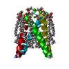 4qk7C 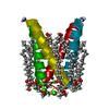 4qkcC  4qklC 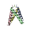 3c9jS  4qk6 C: citing same article ( S: Starting model for refinement |
|---|---|
| Similar structure data |


-Links
 PDBj
PDBj

- Assembly
| Deposited unit | 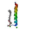
| |||||||||||||||||||||
|---|---|---|---|---|---|---|---|---|---|---|---|---|---|---|---|---|---|---|---|---|---|---|
| 1 | 
| |||||||||||||||||||||
| Unit cell |
| |||||||||||||||||||||
| Components on special symmetry positions |
|
-Components
-Protein/peptide , 1 types, 1 molecules A
| #1: Protein/peptide | Mass: 2754.340 Da / Num. of mol.: 1 / Source method: obtained synthetically Details: This sequence is found in the influenza A virus (strain A/Udorn/307/1972 H3N2) and was manually synthesized using Fmoc chemistry. N- and C-terminal modifications are present. Source: (synth.) Influenza A virus / References: UniProt: W8PGZ1, UniProt: P0DOF5*PLUS |
|---|
-Non-polymers , 5 types, 23 molecules 


| #2: Chemical |  Mass: 40.078 Da / Num. of mol.: 2 / Source method: obtained synthetically / Formula: Ca Mass: 40.078 Da / Num. of mol.: 2 / Source method: obtained synthetically / Formula: Ca#3: Chemical | ChemComp-CL / | Chloride Mass: 35.453 Da / Num. of mol.: 1 / Source method: obtained synthetically / Formula: Cl Mass: 35.453 Da / Num. of mol.: 1 / Source method: obtained synthetically / Formula: Cl#4: Chemical | ChemComp-EDO / | Ethylene glycol Mass: 62.068 Da / Num. of mol.: 1 / Source method: obtained synthetically / Formula: C2H6O2 Mass: 62.068 Da / Num. of mol.: 1 / Source method: obtained synthetically / Formula: C2H6O2#5: Chemical | ChemComp-OLB / ( |  Mass: 356.540 Da / Num. of mol.: 1 / Source method: obtained synthetically / Formula: C21H40O4 Mass: 356.540 Da / Num. of mol.: 1 / Source method: obtained synthetically / Formula: C21H40O4#6: Water | ChemComp-HOH / | WaterMass: 18.015 Da / Num. of mol.: 18 / Source method: isolated from a natural source / Formula: H2O |
|---|
-Experimental details
-Experiment
| Experiment | Method: X-RAY DIFFRACTION / Number of used crystals: 1 |
|---|
- Sample preparation
| Crystal | Density Matthews: 2.77 Å3/Da / Density % sol: 55.58 % |
|---|---|
| Crystal grow | Temperature: 293 K / Method: lcp sandwich plates / pH: 5.5 Details: 1.8 M calcium chloride, 0.9 M MES pH 5.5, 39.6% v/v PEG 400, 0.01M MnCl2, 4H2O additive, LCP sandwich plates, temperature 293K |
-Data collection
| Diffraction | Mean temperature: 273 K |
|---|---|
| Diffraction source | Source: SYNCHROTRON / Site: ALS  / Beamline: 8.3.1 / Wavelength: 1.115869 Å / Beamline: 8.3.1 / Wavelength: 1.115869 Å |
| Detector | Type: ADSC QUANTUM 315r / Detector: CCD / Date: Nov 23, 2013 |
| Radiation | Monochromator: Double flat crystal, Si(111) / Protocol: SINGLE WAVELENGTH / Monochromatic (M) / Laue (L): M / Scattering type: x-ray |
| Radiation wavelength | Wavelength: 1.115869 Å / Relative weight: 1 |
| Reflection | Resolution: 1.44→27.48 Å / Num. all: 5433 / Num. obs: 5254 / % possible obs: 96.7 % / Observed criterion σ(F): 0 / Observed criterion σ(I): 1.4 / Redundancy: 2.4 % / Biso Wilson estimate: 14.85 Å2 / Rmerge(I) obs: 0.048 / Net I/σ(I): 11.4 |
| Reflection shell | Resolution: 1.44→1.47 Å / Redundancy: 2.4 % / Rmerge(I) obs: 0.531 / Mean I/σ(I) obs: 1.4 / % possible all: 98 |
- Processing
| Software |
| |||||||||||||||||||||||||||||||||||
|---|---|---|---|---|---|---|---|---|---|---|---|---|---|---|---|---|---|---|---|---|---|---|---|---|---|---|---|---|---|---|---|---|---|---|---|---|
| Refinement | Method to determine structure: MOLECULAR REPLACEMENT Starting model: chain A of 3C9J Resolution: 1.44→18.001 Å / SU ML: 0.17 / σ(F): 1.4 / Phase error: 22.73 / Stereochemistry target values: ML
| |||||||||||||||||||||||||||||||||||
| Solvent computation | Shrinkage radii: 0.9 Å / VDW probe radii: 1.11 Å / Solvent model: FLAT BULK SOLVENT MODEL | |||||||||||||||||||||||||||||||||||
| Refinement step | Cycle: LAST / Resolution: 1.44→18.001 Å
| |||||||||||||||||||||||||||||||||||
| Refine LS restraints |
| |||||||||||||||||||||||||||||||||||
| LS refinement shell |
|
