Resolution: 1.37→49.32 Å / Cor.coef. Fo:Fc: 0.971 / Cor.coef. Fo:Fc free: 0.96 / SU B: 2.845 / SU ML: 0.053 / Cross valid method: THROUGHOUT / ESU R: 0.061 / ESU R Free: 0.068 / Stereochemistry target values: MAXIMUM LIKELIHOOD / Details: HYDROGENS HAVE BEEN ADDED IN THE RIDING POSITIONS
Rfactor
Num. reflection
% reflection
Selection details
Rfree
0.22029
2265
5.1 %
RANDOM
Rwork
0.17915
-
-
-
obs
0.18128
41955
96.73 %
-
all
-
43373
-
-
Solvent computation
Ion probe radii: 0.8 Å / Shrinkage radii: 0.8 Å / VDW probe radii: 1.2 Å / Solvent model: MASK
Displacement parameters
Biso mean: 26.268 Å2
Baniso -1
Baniso -2
Baniso -3
1-
0.44 Å2
-0 Å2
-1.28 Å2
2-
-
-0.46 Å2
-0 Å2
3-
-
-
0.24 Å2
Refinement step
Cycle: LAST / Resolution: 1.37→49.32 Å
Protein
Nucleic acid
Ligand
Solvent
Total
Num. atoms
1706
0
59
232
1997
Refine LS restraints
Refine-ID
Type
Dev ideal
Dev ideal target
Number
X-RAY DIFFRACTION
r_bond_refined_d
0.026
0.019
1791
X-RAY DIFFRACTION
r_bond_other_d
0.001
0.02
1727
X-RAY DIFFRACTION
r_angle_refined_deg
2.51
2.035
2421
X-RAY DIFFRACTION
r_angle_other_deg
1.357
3
3986
X-RAY DIFFRACTION
r_dihedral_angle_1_deg
5.54
5
216
X-RAY DIFFRACTION
r_dihedral_angle_2_deg
33.683
24.268
82
X-RAY DIFFRACTION
r_dihedral_angle_3_deg
12.297
15
335
X-RAY DIFFRACTION
r_dihedral_angle_4_deg
17.163
15
16
X-RAY DIFFRACTION
r_chiral_restr
0.151
0.2
266
X-RAY DIFFRACTION
r_gen_planes_refined
0.015
0.021
1969
X-RAY DIFFRACTION
r_gen_planes_other
0.008
0.02
377
X-RAY DIFFRACTION
r_mcbond_it
1.499
1.394
867
X-RAY DIFFRACTION
r_mcbond_other
1.496
1.391
866
X-RAY DIFFRACTION
r_mcangle_it
1.88
2.096
1082
X-RAY DIFFRACTION
r_mcangle_other
1.882
2.098
1083
X-RAY DIFFRACTION
r_scbond_it
3.07
1.719
924
X-RAY DIFFRACTION
r_scbond_other
3.068
1.718
925
X-RAY DIFFRACTION
r_scangle_other
4.615
2.421
1340
X-RAY DIFFRACTION
r_long_range_B_refined
8.48
13.597
2244
X-RAY DIFFRACTION
r_long_range_B_other
8.372
12.373
2139
LS refinement shell
Resolution: 1.371→1.407 Å / Total num. of bins used: 20
Rfactor
Num. reflection
% reflection
Rfree
0.353
152
-
Rwork
0.318
2959
-
obs
-
-
92.31 %
Refinement TLS params.
Method: refined / Refine-ID: X-RAY DIFFRACTION
ID
L11 (°2)
L12 (°2)
L13 (°2)
L22 (°2)
L23 (°2)
L33 (°2)
S11 (Å °)
S12 (Å °)
S13 (Å °)
S21 (Å °)
S22 (Å °)
S23 (Å °)
S31 (Å °)
S32 (Å °)
S33 (Å °)
T11 (Å2)
T12 (Å2)
T13 (Å2)
T22 (Å2)
T23 (Å2)
T33 (Å2)
Origin x (Å)
Origin y (Å)
Origin z (Å)
1
2.8741
0
-0.012
2.5501
0.2082
2.7577
-0.0182
0.2846
0.0287
-0.2994
0.0971
-0.0391
-0.0579
0.17
-0.0788
0.0398
-0.0249
0.0048
0.0776
-0.0121
0.0122
21.8059
-1.4839
-3.7486
2
4.2256
-0.3053
1.4702
0.7974
-0.141
1.9854
-0.018
-0.0356
-0.0549
-0.0044
0.034
0.062
-0.1705
-0.014
-0.016
0.0522
0.0421
-0.0164
0.0604
-0.035
0.0668
7.6959
-15.8477
-6.3669
3
1.6299
-0.0506
0.2372
1.3028
0.2538
1.5891
0.0979
-0.101
-0.1013
0.1719
0.0095
0.054
0.0837
0.0198
-0.1075
0.0316
-0.0088
-0.0054
0.0283
0.0046
0.0409
18.2193
-6.9182
14.3104
4
3.2239
3.4441
-1.8329
4.3381
-1.5478
1.829
-0.0821
0.1082
-0.1499
-0.1786
0.0548
-0.027
0.2763
0.0369
0.0274
0.2055
0.0275
-0.049
0.1576
-0.0434
0.2045
16.918
-11.3787
5.0815
Refinement TLS group
ID
Refine-ID
Refine TLS-ID
Auth asym-ID
Auth seq-ID
1
X-RAY DIFFRACTION
1
B
31 - 60
2
X-RAY DIFFRACTION
2
B
127 - 164
3
X-RAY DIFFRACTION
3
B
1 - 30
4
X-RAY DIFFRACTION
3
B
61 - 126
5
X-RAY DIFFRACTION
3
B
165 - 217
6
X-RAY DIFFRACTION
4
B
302
+
About Yorodumi
-
News
-
Feb 9, 2022. New format data for meta-information of EMDB entries
New format data for meta-information of EMDB entries
Version 3 of the EMDB header file is now the official format.
The previous official version 1.9 will be removed from the archive.
In the structure databanks used in Yorodumi, some data are registered as the other names, "COVID-19 virus" and "2019-nCoV". Here are the details of the virus and the list of structure data.
Jan 31, 2019. EMDB accession codes are about to change! (news from PDBe EMDB page)
EMDB accession codes are about to change! (news from PDBe EMDB page)
The allocation of 4 digits for EMDB accession codes will soon come to an end. Whilst these codes will remain in use, new EMDB accession codes will include an additional digit and will expand incrementally as the available range of codes is exhausted. The current 4-digit format prefixed with “EMD-” (i.e. EMD-XXXX) will advance to a 5-digit format (i.e. EMD-XXXXX), and so on. It is currently estimated that the 4-digit codes will be depleted around Spring 2019, at which point the 5-digit format will come into force.
The EM Navigator/Yorodumi systems omit the EMD- prefix.
Related info.:Q: What is EMD? / ID/Accession-code notation in Yorodumi/EM Navigator
Yorodumi is a browser for structure data from EMDB, PDB, SASBDB, etc.
This page is also the successor to EM Navigator detail page, and also detail information page/front-end page for Omokage search.
The word "yorodu" (or yorozu) is an old Japanese word meaning "ten thousand". "mi" (miru) is to see.
Related info.:EMDB / PDB / SASBDB / Comparison of 3 databanks / Yorodumi Search / Aug 31, 2016. New EM Navigator & Yorodumi / Yorodumi Papers / Jmol/JSmol / Function and homology information / Changes in new EM Navigator and Yorodumi
 Movie
Movie Controller


 Open data
 Basic information
 Components
 Keywords
 Function and homology information
 Authors
 Citation
 Structure visualization
 Downloads & links
 Other downloads
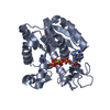


 PDBj
PDBj
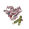
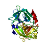
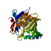
 Assembly
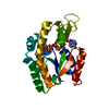

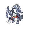
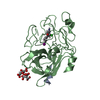
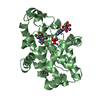
 Mass: 65.409 Da / Num. of mol.: 1 / Source method: obtained synthetically / Formula: Zn
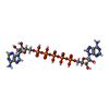
 Mass: 916.367 Da / Num. of mol.: 1 / Source method: obtained synthetically / Formula: C20H29N10O22P5
 Mass: 24.305 Da / Num. of mol.: 1 / Source method: obtained synthetically / Formula: Mg
 Mass: 18.015 Da / Num. of mol.: 232 / Source method: isolated from a natural source / Formula: H2O
 Sample preparation
 / Beamline: 7A (6B, 6C1) / Wavelength: 0.9795 Å
 Processing