 Movie
Movie Controller

[English] 日本語
 Yorodumi
- PDB-4q6j: Crystal Structure of EAL domain Protein from Listeria monocytogen... -
+ Open data

- Basic information
| Entry | Database: PDB / ID: 4q6j | ||||||
|---|---|---|---|---|---|---|---|
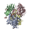
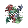
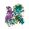
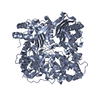
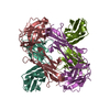
| Title | Crystal Structure of EAL domain Protein from Listeria monocytogenes EGD-e | ||||||
 Components Components | Lmo0131 protein | ||||||
 Keywords Keywords | UNKNOWN FUNCTION /  Structural Genomics / PSI-Biology / Midwest Center for Structural Genomics / MCSG / TIM barrel / alpha-betafold Structural Genomics / PSI-Biology / Midwest Center for Structural Genomics / MCSG / TIM barrel / alpha-betafold | ||||||
| Function / homology |  Function and homology informationEAL domain / Putative diguanylate phosphodiesterase / EAL domain / EAL domain superfamily / EAL domain / EAL domain profile. / TIM Barrel / Alpha-Beta Barrel / Alpha Beta Function and homology informationEAL domain / Putative diguanylate phosphodiesterase / EAL domain / EAL domain superfamily / EAL domain / EAL domain profile. / TIM Barrel / Alpha-Beta Barrel / Alpha BetaSimilarity search - Domain/homology | ||||||
| Biological species |  Listeria monocytogenes (bacteria) Listeria monocytogenes (bacteria) | ||||||
| Method | X-RAY DIFFRACTION / SYNCHROTRON / SAD / Resolution: 1.369 Å | ||||||
 Authors Authors | Kim, Y. / Bigelow, L. / Clancy, S. / Joachimiak, A. / Midwest Center for Structural Genomics (MCSG) | ||||||
 Citation Citation | Journal: To be Published Title: Crystal Structure of EAL domain Protein from Listeria monocytogenes EGD-e Authors: Kim, Y. / Bigelow, L. / Clancy, S. / Joachimiak, A. / Midwest Center for Structural Genomics (MCSG) | ||||||
| History |
|
- Structure visualization
| Structure viewer | Molecule: MolmilJmol/JSmol |
|---|
- Downloads & links
-Download
| PDBx/mmCIF format | 4q6j.cif.gz | 238.9 KB | Display | PDBx/mmCIF format |
|---|---|---|---|---|
| PDB format | pdb4q6j.ent.gz | 202.7 KB | Display | PDB format |
| PDBx/mmJSON format | 4q6j.json.gz | Tree view | PDBx/mmJSON format | |
| Others |  Other downloads Other downloads |
-Validation report
| Arichive directory | https://data.pdbj.org/pub/pdb/validation_reports/q6/4q6jftp://data.pdbj.org/pub/pdb/validation_reports/q6/4q6j | HTTPS FTP |
|---|
-Related structure data
| Similar structure data | |
|---|---|
| Other databases |


-Links
 PDBj
PDBj

- Assembly
| Deposited unit | 
| ||||||||
|---|---|---|---|---|---|---|---|---|---|
| 1 | 
| ||||||||
| Unit cell |
| ||||||||
| Components on special symmetry positions |
|
-Components
-Protein , 1 types, 2 molecules AB
| #1: Protein | Mass: 29320.643 Da / Num. of mol.: 2 Source method: isolated from a genetically manipulated source Source: (gene. exp.) Listeria monocytogenes (bacteria) / Strain: EGD-e / Gene: lmo0131 / Plasmid: pMCSG73 / Production host: Escherichia coli (E. coli) / Strain (production host): BL21(DE3) gold / References: UniProt: Q8YAJ4 |
|---|
-Non-polymers , 5 types, 410 molecules 


| #2: Chemical | Phosphate Mass: 94.971 Da / Num. of mol.: 2 / Source method: obtained synthetically / Formula: PO4 Mass: 94.971 Da / Num. of mol.: 2 / Source method: obtained synthetically / Formula: PO4#3: Chemical | ChemComp-EDO / Ethylene glycol Mass: 62.068 Da / Num. of mol.: 13 / Source method: obtained synthetically / Formula: C2H6O2 Mass: 62.068 Da / Num. of mol.: 13 / Source method: obtained synthetically / Formula: C2H6O2#4: Chemical | ChemComp-GOL / | Glycerol Mass: 92.094 Da / Num. of mol.: 1 / Source method: obtained synthetically / Formula: C3H8O3 Mass: 92.094 Da / Num. of mol.: 1 / Source method: obtained synthetically / Formula: C3H8O3#5: Chemical | ChemComp-ACY / | Acetic acid Mass: 60.052 Da / Num. of mol.: 1 / Source method: obtained synthetically / Formula: C2H4O2 Mass: 60.052 Da / Num. of mol.: 1 / Source method: obtained synthetically / Formula: C2H4O2#6: Water | ChemComp-HOH / | WaterMass: 18.015 Da / Num. of mol.: 393 / Source method: isolated from a natural source / Formula: H2O |
|---|
-Experimental details
-Experiment
| Experiment | Method: X-RAY DIFFRACTION / Number of used crystals: 1 |
|---|
- Sample preparation
| Crystal | Density Matthews: 2.58 Å3/Da / Density % sol: 52.37 % |
|---|---|
| Crystal grow | Temperature: 289 K / Method: vapor diffusion, sitting drop / pH: 7.5 Details: 0.1 M HEPES pH 7.5, 0.8 M sodium phosphate, 0.8 M postassium phosphate, VAPOR DIFFUSION, SITTING DROP, temperature 289K |
-Data collection
| Diffraction | Mean temperature: 100 K |
|---|---|
| Diffraction source | Source: SYNCHROTRON / Site: APS  / Beamline: 19-ID / Wavelength: 0.97921 Å / Beamline: 19-ID / Wavelength: 0.97921 Å |
| Detector | Type: ADSC QUANTUM 315r / Detector: CCD / Date: Jun 14, 2013 / Details: mirrors |
| Radiation | Monochromator: double crystal monochromator / Protocol: SINGLE WAVELENGTH / Monochromatic (M) / Laue (L): M / Scattering type: x-ray |
| Radiation wavelength | Wavelength: 0.97921 Å / Relative weight: 1 |
| Reflection twin | Operator: -h-2*l,-k,l / Fraction: 0.5 |
| Reflection | Resolution: 1.37→50 Å / Num. all: 120312 / Num. obs: 120312 / % possible obs: 96.2 % / Observed criterion σ(F): 0 / Observed criterion σ(I): 0 / Redundancy: 4.5 % / Biso Wilson estimate: 17.02 Å2 / Rsym value: 0.083 / Net I/σ(I): 13.1 |
| Reflection shell | Resolution: 1.37→1.39 Å / Redundancy: 3.5 % / Mean I/σ(I) obs: 1.8 / Num. unique all: 4456 / Rsym value: 0.6 / % possible all: 72.2 |
- Processing
| Software |
| |||||||||||||||||||||||||||||||||||||||||||||||||||||||||||||||||||||||||||||||||||||||||||||||||||||||||||||||||||||||||||||||||||||||||||||||||||
|---|---|---|---|---|---|---|---|---|---|---|---|---|---|---|---|---|---|---|---|---|---|---|---|---|---|---|---|---|---|---|---|---|---|---|---|---|---|---|---|---|---|---|---|---|---|---|---|---|---|---|---|---|---|---|---|---|---|---|---|---|---|---|---|---|---|---|---|---|---|---|---|---|---|---|---|---|---|---|---|---|---|---|---|---|---|---|---|---|---|---|---|---|---|---|---|---|---|---|---|---|---|---|---|---|---|---|---|---|---|---|---|---|---|---|---|---|---|---|---|---|---|---|---|---|---|---|---|---|---|---|---|---|---|---|---|---|---|---|---|---|---|---|---|---|---|---|---|---|
| Refinement | Method to determine structure: SAD / Resolution: 1.369→30.069 Å / Isotropic thermal model: anisotropic / Cross valid method: THROUGHOUT / σ(F): 0 / Phase error: 18.92 / Stereochemistry target values: TWIN_LSQ_F Details: both Freidel pair reflections were used in refinement, which is reflected the refinement statistics
| |||||||||||||||||||||||||||||||||||||||||||||||||||||||||||||||||||||||||||||||||||||||||||||||||||||||||||||||||||||||||||||||||||||||||||||||||||
| Solvent computation | Shrinkage radii: 0.9 Å / VDW probe radii: 1.11 Å / Solvent model: FLAT BULK SOLVENT MODEL | |||||||||||||||||||||||||||||||||||||||||||||||||||||||||||||||||||||||||||||||||||||||||||||||||||||||||||||||||||||||||||||||||||||||||||||||||||
| Displacement parameters | Biso mean: 21.9 Å2 | |||||||||||||||||||||||||||||||||||||||||||||||||||||||||||||||||||||||||||||||||||||||||||||||||||||||||||||||||||||||||||||||||||||||||||||||||||
| Refinement step | Cycle: LAST / Resolution: 1.369→30.069 Å
| |||||||||||||||||||||||||||||||||||||||||||||||||||||||||||||||||||||||||||||||||||||||||||||||||||||||||||||||||||||||||||||||||||||||||||||||||||
| Refine LS restraints |
| |||||||||||||||||||||||||||||||||||||||||||||||||||||||||||||||||||||||||||||||||||||||||||||||||||||||||||||||||||||||||||||||||||||||||||||||||||
| LS refinement shell | Refine-ID: X-RAY DIFFRACTION
|
