 Movie
Movie Controller

[English] 日本語
 Yorodumi
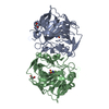
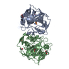
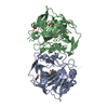
- PDB-4q0l: Crystal structure of catalytic domain of human carbonic anhydrase... -
+ Open data

- Basic information
| Entry | Database: PDB / ID: 4q0l | ||||||
|---|---|---|---|---|---|---|---|
| Title | Crystal structure of catalytic domain of human carbonic anhydrase isozyme XII with inhibitor | ||||||
 Components Components | Carbonic anhydrase 12 | ||||||
 Keywords Keywords | LYASE/LYASE INHIBITOR / drug design / carbonic anhydrase / benzenesulfonamide / metal-binding / lyase-lyase inhibitor comple / LYASE-LYASE INHIBITOR complex | ||||||
| Function / homology |  Function and homology information Function and homology informationchloride ion homeostasis / estrous cycle / Reversible hydration of carbon dioxide / carbonic anhydrase / carbonate dehydratase activity / one-carbon metabolic process / basolateral plasma membrane / apical plasma membrane / zinc ion binding / membrane / plasma membraneSimilarity search - Function | ||||||
| Biological species |  Homo sapiens (human) Homo sapiens (human) | ||||||
| Method | X-RAY DIFFRACTION / molecular replacement / Resolution: 2 Å | ||||||
 Authors Authors | Smirnov, A. / Manakova, E. / Grazulis, S. | ||||||
 Citation Citation | Journal: J.Med.Chem. / Year: 2014 Title: Discovery and characterization of novel selective inhibitors of carbonic anhydrase IX. Authors: Dudutiene, V. / Matuliene, J. / Smirnov, A. / Timm, D.D. / Zubriene, A. / Baranauskiene, L. / Morkunaite, V. / Smirnoviene, J. / Michailoviene, V. / Juozapaitiene, V. / Mickeviciute, A. / ...Authors: Dudutiene, V. / Matuliene, J. / Smirnov, A. / Timm, D.D. / Zubriene, A. / Baranauskiene, L. / Morkunaite, V. / Smirnoviene, J. / Michailoviene, V. / Juozapaitiene, V. / Mickeviciute, A. / Kazokaite, J. / Baksyte, S. / Kasiliauskaite, A. / Jachno, J. / Revuckiene, J. / Kisonaite, M. / Pilipuityte, V. / Ivanauskaite, E. / Milinaviciute, G. / Smirnovas, V. / Petrikaite, V. / Kairys, V. / Petrauskas, V. / Norvaisas, P. / Linge, D. / Gibieza, P. / Capkauskaite, E. / Zaksauskas, A. / Kazlauskas, E. / Manakova, E. / Grazulis, S. / Ladbury, J.E. / Matulis, D. | ||||||
| History |
|
- Structure visualization
| Structure viewer | Molecule: MolmilJmol/JSmol |
|---|
- Downloads & links
-Download
| PDBx/mmCIF format | 4q0l.cif.gz | 226.5 KB | Display | PDBx/mmCIF format |
|---|---|---|---|---|
| PDB format | pdb4q0l.ent.gz | 181.9 KB | Display | PDB format |
| PDBx/mmJSON format | 4q0l.json.gz | Tree view | PDBx/mmJSON format | |
| Others |  Other downloads Other downloads |
-Validation report
| Arichive directory | https://data.pdbj.org/pub/pdb/validation_reports/q0/4q0lftp://data.pdbj.org/pub/pdb/validation_reports/q0/4q0l | HTTPS FTP |
|---|
-Related structure data
| Related structure data |  4pyxC  4pyyC  4pzhC  4q06C  4q07C  4q08C  4q09C 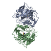 1jd0S C: citing same article ( S: Starting model for refinement |
|---|---|
| Similar structure data |

-Links
 PDBj
PDBj
- Assembly
| Deposited unit | 
| ||||||||
|---|---|---|---|---|---|---|---|---|---|
| 1 | 
| ||||||||
| 2 | 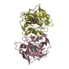
| ||||||||
| 3 | 
| ||||||||
| 4 | 
| ||||||||
| 5 | 
| ||||||||
| 6 | 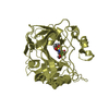
| ||||||||
| Unit cell |
| ||||||||
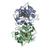
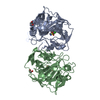
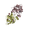
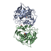
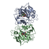
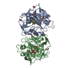
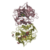
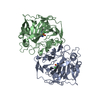
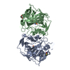
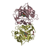
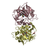
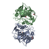
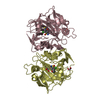
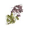
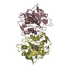
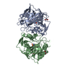
| Details | The asymmetric unit contains two dimers: first dimer (A, B chain) and second dimer (C, D chain). |
-Components
| #1: Protein | / Carbonate dehydratase XII / Carbonic anhydrase XII / CA-XII / Tumor antigen HOM-RCC-3.1.3 Mass: 29917.318 Da / Num. of mol.: 4 Source method: isolated from a genetically manipulated source Source: (gene. exp.) Homo sapiens (human) / Gene: CA12 / Plasmid: pET21a / Production host:  Escherichia coli (E. coli) / Strain (production host): Rosetta(DE3) / References: UniProt: O43570, carbonic anhydrase Escherichia coli (E. coli) / Strain (production host): Rosetta(DE3) / References: UniProt: O43570, carbonic anhydrase#2: Chemical | ChemComp-ZN /   Mass: 65.409 Da / Num. of mol.: 4 / Source method: obtained synthetically / Formula: Zn Mass: 65.409 Da / Num. of mol.: 4 / Source method: obtained synthetically / Formula: Zn#3: Chemical | ChemComp-V14 /   Mass: 444.489 Da / Num. of mol.: 4 / Source method: obtained synthetically / Formula: C16H23F3N2O5S2 Mass: 444.489 Da / Num. of mol.: 4 / Source method: obtained synthetically / Formula: C16H23F3N2O5S2#4: Water | ChemComp-HOH / | Water Mass: 18.015 Da / Num. of mol.: 320 / Source method: isolated from a natural source / Formula: H2O Mass: 18.015 Da / Num. of mol.: 320 / Source method: isolated from a natural source / Formula: H2O |
|---|
-Experimental details
-Experiment
| Experiment | Method: X-RAY DIFFRACTION / Number of used crystals: 1 |
|---|
- Sample preparation
| Crystal | Density Matthews: 1.92 Å3/Da / Density % sol: 35.4 % |
|---|---|
| Crystal grow | Temperature: 291 K / Method: vapor diffusion, sitting drop / pH: 5 Details: Crystallization buffer: 0.1M ammonium citrate with pH 5.0 and 18% of PEG4000, VAPOR DIFFUSION, SITTING DROP, temperature 291K |
-Data collection
| Diffraction | Mean temperature: 100 K | ||||||||||||||||||||||||||||||||||||||||||||||||||||||||||||||||||||||||||||||||||||||||
|---|---|---|---|---|---|---|---|---|---|---|---|---|---|---|---|---|---|---|---|---|---|---|---|---|---|---|---|---|---|---|---|---|---|---|---|---|---|---|---|---|---|---|---|---|---|---|---|---|---|---|---|---|---|---|---|---|---|---|---|---|---|---|---|---|---|---|---|---|---|---|---|---|---|---|---|---|---|---|---|---|---|---|---|---|---|---|---|---|---|
| Diffraction source | Source: ROTATING ANODE / Type: RIGAKU MICROMAX-007 HF / Wavelength: 1.5418 Å | ||||||||||||||||||||||||||||||||||||||||||||||||||||||||||||||||||||||||||||||||||||||||
| Detector | Type: RIGAKU RAXIS IV++ / Detector: IMAGE PLATE / Date: May 15, 2012 | ||||||||||||||||||||||||||||||||||||||||||||||||||||||||||||||||||||||||||||||||||||||||
| Radiation | Monochromator: VARIMAX-HF MIRROR / Protocol: SINGLE WAVELENGTH / Monochromatic (M) / Laue (L): M / Scattering type: x-ray | ||||||||||||||||||||||||||||||||||||||||||||||||||||||||||||||||||||||||||||||||||||||||
| Radiation wavelength | Wavelength: 1.5418 Å / Relative weight: 1 | ||||||||||||||||||||||||||||||||||||||||||||||||||||||||||||||||||||||||||||||||||||||||
| Reflection | Resolution: 2→22.11 Å / Num. obs: 44882 / % possible obs: 74.5 % / Observed criterion σ(F): 0 / Observed criterion σ(I): 0 / Redundancy: 2.9 % / Biso Wilson estimate: 23.407 Å2 / Rmerge(I) obs: 0.068 / Rsym value: 0.068 / Net I/σ(I): 12.2 | ||||||||||||||||||||||||||||||||||||||||||||||||||||||||||||||||||||||||||||||||||||||||
| Reflection shell | Diffraction-ID: 1
|
-Phasing
| Phasing | Method: molecular replacement |
|---|
- Processing
| Software |
| |||||||||||||||||||||||||||||||||||||||||||||
|---|---|---|---|---|---|---|---|---|---|---|---|---|---|---|---|---|---|---|---|---|---|---|---|---|---|---|---|---|---|---|---|---|---|---|---|---|---|---|---|---|---|---|---|---|---|---|
| Refinement | Method to determine structure: molecular replacement Starting model: PDB ENTRY 1JD0 Resolution: 2→22.09 Å / Cor.coef. Fo:Fc: 0.932 / Cor.coef. Fo:Fc free: 0.894 / WRfactor Rfree: 0.327 / WRfactor Rwork: 0.266 / Occupancy max: 1 / Occupancy min: 0.01 / FOM work R set: 0.603 / SU R Cruickshank DPI: 0.824 / SU Rfree: 0.352 / Cross valid method: THROUGHOUT / σ(F): 0 / ESU R: 0.824 / ESU R Free: 0.352 / Stereochemistry target values: MAXIMUM LIKELIHOOD Details: HYDROGENS HAVE BEEN USED IF PRESENT IN THE INPUT U VALUES : REFINED INDIVIDUALLY
| |||||||||||||||||||||||||||||||||||||||||||||
| Solvent computation | Ion probe radii: 0.8 Å / Shrinkage radii: 0.8 Å / VDW probe radii: 1.4 Å / Solvent model: MASK | |||||||||||||||||||||||||||||||||||||||||||||
| Displacement parameters | Biso max: 63.97 Å2 / Biso mean: 20.194 Å2 / Biso min: 5.29 Å2
| |||||||||||||||||||||||||||||||||||||||||||||
| Refinement step | Cycle: LAST / Resolution: 2→22.09 Å
| |||||||||||||||||||||||||||||||||||||||||||||
| Refine LS restraints |
| |||||||||||||||||||||||||||||||||||||||||||||
| LS refinement shell | Resolution: 2→2.052 Å / Total num. of bins used: 20
|
