 Movie
Movie Controller


+ Open data

- Basic information
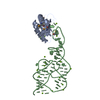
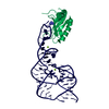
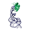
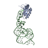
| Entry | Database: PDB / ID: 4prf | |||||||||
|---|---|---|---|---|---|---|---|---|---|---|
| Title | A Second Look at the HDV Ribozyme Structure and Dynamics. | |||||||||
 Components Components |
| |||||||||
 Keywords Keywords | RNA BINDING PROTEIN/RNA / HDV /  ribozyme / RNA / U1A / precursor / TRANSLATION-RNA COMPLEX / RNA BINDING PROTEIN-RNA complex ribozyme / RNA / U1A / precursor / TRANSLATION-RNA COMPLEX / RNA BINDING PROTEIN-RNA complex | |||||||||
| Function / homology |  Function and homology information Function and homology informationU1 snRNP binding / U1 snRNP / U1 snRNA binding / U4/U6 x U5 tri-snRNP complex / mRNA Splicing - Major Pathway / spliceosomal complex / mRNA splicing, via spliceosome / DNA binding / RNA binding / nucleoplasm ...U1 snRNP binding / U1 snRNP / U1 snRNA binding / U4/U6 x U5 tri-snRNP complex / mRNA Splicing - Major Pathway / spliceosomal complex / mRNA splicing, via spliceosome / DNA binding / RNA binding / nucleoplasm / identical protein binding / nucleusSimilarity search - Function | |||||||||
| Biological species |  Homo sapiens (human) Homo sapiens (human) | |||||||||
| Method | X-RAY DIFFRACTION / SYNCHROTRON / MAD / Resolution: 2.395 Å | |||||||||
 Authors Authors | Kapral, G.J. / Jain, S. / Noeske, J. / Doudna, J.A. / Richardson, D.C. / Richardson, J.S. | |||||||||
 Citation Citation | Journal: Nucleic Acids Res. / Year: 2014 Title: New tools provide a second look at HDV ribozyme structure, dynamics and cleavage. Authors: Kapral, G.J. / Jain, S. / Noeske, J. / Doudna, J.A. / Richardson, D.C. / Richardson, J.S. | |||||||||
| History |
|
- Structure visualization
| Structure viewer | Molecule: MolmilJmol/JSmol |
|---|
- Downloads & links
-Download
| PDBx/mmCIF format | 4prf.cif.gz | 169.3 KB | Display | PDBx/mmCIF format |
|---|---|---|---|---|
| PDB format | pdb4prf.ent.gz | 135.7 KB | Display | PDB format |
| PDBx/mmJSON format | 4prf.json.gz | Tree view | PDBx/mmJSON format | |
| Others |  Other downloads Other downloads |
-Validation report
| Arichive directory | https://data.pdbj.org/pub/pdb/validation_reports/pr/4prfftp://data.pdbj.org/pub/pdb/validation_reports/pr/4prf | HTTPS FTP |
|---|
-Related structure data


-Links
 PDBj
PDBj


- Assembly
| Deposited unit | 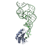
| ||||||||
|---|---|---|---|---|---|---|---|---|---|
| 1 |
| ||||||||
| Unit cell |
| ||||||||
| Components on special symmetry positions |
|
-Components
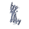
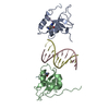
| #1: RNA chain | Mass: 24467.521 Da / Num. of mol.: 1 / Source method: obtained synthetically Details: RNA occurs from Hapatitis Delta Virus pathogen, in vitro transcription with pUc19 | ||
|---|---|---|---|
| #2: Protein | Mass: 11498.472 Da / Num. of mol.: 1 / Fragment: U1A_RBD, UNP residues 98-173 / Mutation: Y31H, Q36R Source method: isolated from a genetically manipulated source Source: (gene. exp.) Homo sapiens (human) / Gene: SNRPA / Plasmid: pET / Production host:  Escherichia coli (E. coli) / References: UniProt: P09012 Escherichia coli (E. coli) / References: UniProt: P09012 | ||
| #3: Chemical | Strontium  Mass: 87.620 Da / Num. of mol.: 2 / Source method: obtained synthetically / Formula: Sr Mass: 87.620 Da / Num. of mol.: 2 / Source method: obtained synthetically / Formula: Sr#4: Water | ChemComp-HOH / | Water Mass: 18.015 Da / Num. of mol.: 42 / Source method: isolated from a natural source / Formula: H2O Mass: 18.015 Da / Num. of mol.: 42 / Source method: isolated from a natural source / Formula: H2O |
-Experimental details
-Experiment
| Experiment | Method: X-RAY DIFFRACTION / Number of used crystals: 1 |
|---|
- Sample preparation
| Crystal | Density Matthews: 3.04 Å3/Da / Density % sol: 59.6 % |
|---|---|
| Crystal grow | Temperature: 277 K / Method: vapor diffusion, hanging drop / pH: 6 Details: SrCl2, NaCl, MPD, Sodium Cacodylate, Spermine-HCl, pH 6.0, VAPOR DIFFUSION, HANGING DROP, temperature 277K |
-Data collection
| Diffraction | Mean temperature: 80 K | |||||||||||||||
|---|---|---|---|---|---|---|---|---|---|---|---|---|---|---|---|---|
| Diffraction source | Source: SYNCHROTRON / Site: ALS  / Beamline: 8.2.1 / Wavelength: 1.0781, 0.9787, 0.9795, 0.9797 / Beamline: 8.2.1 / Wavelength: 1.0781, 0.9787, 0.9795, 0.9797 | |||||||||||||||
| Detector | Type: ADSC QUANTUM 4 / Detector: CCD / Date: Jan 16, 2003 | |||||||||||||||
| Radiation | Monochromator: graphite / Protocol: MAD / Monochromatic (M) / Laue (L): M / Scattering type: x-ray | |||||||||||||||
| Radiation wavelength |
| |||||||||||||||
| Reflection | Resolution: 2.395→50 Å / Num. all: 17315 / % possible obs: 93.1 % / Observed criterion σ(F): 2 / Observed criterion σ(I): 2.56 | |||||||||||||||
| Reflection shell | Resolution: 2.4→2.46 Å / % possible all: 75 |
- Processing
| Software |
| |||||||||||||||||||||||||||||||||||||||||||||||||||||||||||||||||||||||||||
|---|---|---|---|---|---|---|---|---|---|---|---|---|---|---|---|---|---|---|---|---|---|---|---|---|---|---|---|---|---|---|---|---|---|---|---|---|---|---|---|---|---|---|---|---|---|---|---|---|---|---|---|---|---|---|---|---|---|---|---|---|---|---|---|---|---|---|---|---|---|---|---|---|---|---|---|---|
| Refinement | Method to determine structure: MAD / Resolution: 2.395→35.528 Å / SU ML: 0.36 / Cross valid method: THROUGHOUT / σ(F): 1.48 / Phase error: 28.91 / Stereochemistry target values: ML
| |||||||||||||||||||||||||||||||||||||||||||||||||||||||||||||||||||||||||||
| Solvent computation | Shrinkage radii: 0.9 Å / VDW probe radii: 1.11 Å / Solvent model: FLAT BULK SOLVENT MODEL | |||||||||||||||||||||||||||||||||||||||||||||||||||||||||||||||||||||||||||
| Refinement step | Cycle: LAST / Resolution: 2.395→35.528 Å
| |||||||||||||||||||||||||||||||||||||||||||||||||||||||||||||||||||||||||||
| Refine LS restraints |
| |||||||||||||||||||||||||||||||||||||||||||||||||||||||||||||||||||||||||||
| LS refinement shell |
| |||||||||||||||||||||||||||||||||||||||||||||||||||||||||||||||||||||||||||
| Refinement TLS params. | Method: refined / Refine-ID: X-RAY DIFFRACTION
| |||||||||||||||||||||||||||||||||||||||||||||||||||||||||||||||||||||||||||
| Refinement TLS group |
|
