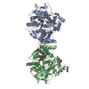
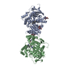
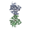
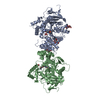
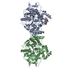
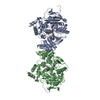
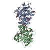
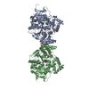
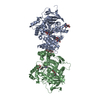
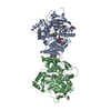
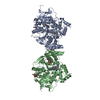
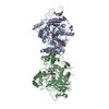
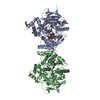
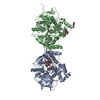
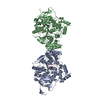
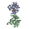
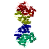
Entry Database : PDB / ID : 4pqeTitle Crystal Structure of Human Acetylcholinesterase Acetylcholinesterase Keywords / / / / Function / homology Function Domain/homology Component
/ / / / / / / / / / / / / / / / / / / / / / / / / / / / / / / / / / / / / / / / / / / / / / / / / / / / / / / / / / / / / / / / / / / / / / / Biological species Homo sapiens (human)Method / / / Resolution : 2.9 Å Authors Dym, O. / Unger, T. / Toker, L. / Silman, I. / Sussman, J.L. / Israel Structural Proteomics Center (ISPC) Journal : To be Published Title : Crystal Structure of Human AcetylcholinesteraseAuthors : Dym, O. / Unger, T. / Toker, L. / Silman, I. / Sussman, J.L. / Israel Structural Proteomics Center (ISPC) History Deposition Mar 2, 2014 Deposition site / Processing site Revision 1.0 Apr 8, 2015 Provider / Type Revision 1.1 Sep 20, 2023 Group Data collection / Database references ... Data collection / Database references / Derived calculations / Refinement description Category chem_comp_atom / chem_comp_bond ... chem_comp_atom / chem_comp_bond / database_2 / pdbx_initial_refinement_model / struct_conn Item _database_2.pdbx_DOI / _database_2.pdbx_database_accession ... _database_2.pdbx_DOI / _database_2.pdbx_database_accession / _struct_conn.ptnr1_auth_seq_id / _struct_conn.ptnr1_label_seq_id / _struct_conn.ptnr2_auth_seq_id / _struct_conn.ptnr2_label_seq_id
Show all Show less
 Movie
Movie Controller


 Open data
 Basic information
 Components
 Keywords
 Function and homology information
 Authors
 Citation
 Structure visualization
 Downloads & links
 Other downloads
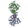

 PDBj
PDBj


 Assembly
 Mass: 18.015 Da / Num. of mol.: 12 / Source method: isolated from a natural source / Formula: H2O
 Sample preparation
 / Beamline: ID23-2 / Wavelength: 0.8726 Å
 Processing