[English] 日本語
Yorodumi
- PDB-4ph1: C-terminal domain of capsid protein from bovine leukemia virus -

+
Open data


ID or keywords:

Loading...

-
Basic information

Entry
Database: PDB / ID: 4ph1
TitleC-terminal domain of capsid protein from bovine leukemia virus
ComponentsBLV capsid
Keywords
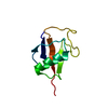
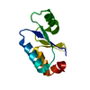
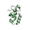
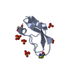
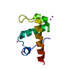
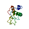
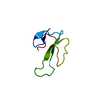
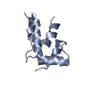
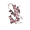
VIRAL PROTEIN / retroviral capsid CTD / all alpha
Function / homology
Function and homology information


DNA integration / viral genome integration into host DNA / establishment of integrated proviral latency / RNA-directed DNA polymerase activity / RNA-DNA hybrid ribonuclease activity / viral nucleocapsid / DNA recombination / aspartic-type endopeptidase activity / symbiont entry into host cell / structural molecule activity ...DNA integration / viral genome integration into host DNA / establishment of integrated proviral latency / RNA-directed DNA polymerase activity / RNA-DNA hybrid ribonuclease activity / viral nucleocapsid / DNA recombination / aspartic-type endopeptidase activity / symbiont entry into host cell / structural molecule activity / proteolysis / DNA binding / zinc ion binding
Similarity search - Function
Delta-retroviral matrix protein / Major core protein p19 / Retrovirus capsid C-terminal domain / Non-ribosomal Peptide Synthetase Peptidyl Carrier Protein; Chain A / gag protein p24 N-terminal domain / Integrase DNA binding domain / Integrase, C-terminal, retroviral / Integrase DNA binding domain profile. / RNase H / Integrase core domain ...Delta-retroviral matrix protein / Major core protein p19 / Retrovirus capsid C-terminal domain / Non-ribosomal Peptide Synthetase Peptidyl Carrier Protein; Chain A / gag protein p24 N-terminal domain / Integrase DNA binding domain / Integrase, C-terminal, retroviral / Integrase DNA binding domain profile. / RNase H / Integrase core domain / Integrase, catalytic core / Integrase catalytic domain profile. / Retroviral nucleocapsid Gag protein p24, C-terminal domain / Gag protein p24 C-terminal domain / Ribonuclease H domain / RNase H type-1 domain profile. / Reverse transcriptase (RNA-dependent DNA polymerase) / Reverse transcriptase domain / Reverse transcriptase (RT) catalytic domain profile. / Retropepsins / Retroviral aspartyl protease / Aspartyl protease, retroviral-type family profile. / Peptidase A2A, retrovirus, catalytic / Retrovirus capsid, C-terminal / Retroviral matrix protein / Retrovirus capsid, N-terminal / zinc finger / Zinc knuckle / Zinc finger, CCHC-type superfamily / Zinc finger, CCHC-type / Zinc finger CCHC-type profile. / Ribonuclease H superfamily / Aspartic peptidase domain superfamily / Ribonuclease H-like superfamily / Reverse transcriptase/Diguanylate cyclase domain / DNA/RNA polymerase superfamily / Orthogonal Bundle / Mainly Alpha
Similarity search - Domain/homology
Gag-Pro-Pol polyprotein
Similarity search - Component
Biological speciesBovine leukemia virus
MethodX-RAY DIFFRACTION / MOLECULAR REPLACEMENT / Resolution: 2.46 Å
Model detailsNTD lacking the N-term beta-hairpin
AuthorsTrajtenberg, F. / Obal, G. / Pritsch, O. / Buschiazzo, A.
CitationJournal: Science / Year: 2015
Title: STRUCTURAL VIROLOGY. Conformational plasticity of a native retroviral capsid revealed by x-ray crystallography.
Authors: Obal, G. / Trajtenberg, F. / Carrion, F. / Tome, L. / Larrieux, N. / Zhang, X. / Pritsch, O. / Buschiazzo, A.
History
DepositionMay 3, 2014Deposition site: RCSB / Processing site: RCSB
Revision 1.0Jun 10, 2015Provider: repository / Type: Initial release
Revision 1.1Jun 17, 2015Group: Database references
Revision 1.2Jul 15, 2015Group: Database references
Revision 1.3Nov 22, 2017Group: Database references / Derived calculations ...Database references / Derived calculations / Other / Refinement description / Source and taxonomy / Structure summary
Category: citation / entity_src_gen ...citation / entity_src_gen / pdbx_database_status / pdbx_struct_assembly / pdbx_struct_oper_list / software / struct_keywords
Item: _citation.journal_id_CSD / _entity_src_gen.pdbx_alt_source_flag ..._citation.journal_id_CSD / _entity_src_gen.pdbx_alt_source_flag / _pdbx_database_status.pdb_format_compatible / _pdbx_struct_assembly.oligomeric_details / _pdbx_struct_oper_list.symmetry_operation / _software.classification / _struct_keywords.text
Revision 1.4Sep 27, 2023Group: Data collection / Database references / Refinement description
Category: chem_comp_atom / chem_comp_bond ...chem_comp_atom / chem_comp_bond / database_2 / pdbx_initial_refinement_model / refine_hist
Item: _database_2.pdbx_DOI / _database_2.pdbx_database_accession ..._database_2.pdbx_DOI / _database_2.pdbx_database_accession / _refine_hist.pdbx_number_atoms_nucleic_acid / _refine_hist.pdbx_number_atoms_protein

-
Structure visualization

Structure viewerMolecule:
MolmilJmol/JSmol

Downloads & links

-
Assembly

Deposited unit
A: BLV capsid
B: BLV capsid
C: BLV capsid


Theoretical massNumber of molelcules
Total (without water)29,9143
Polymers29,9143
Non-polymers00
Water1,65792
1
A: BLV capsid


Theoretical massNumber of molelcules
Total (without water)9,9711
Polymers9,9711
Non-polymers00
Water181
TypeNameSymmetry operationNumber
identity operation1_555x,y,z1
2
B: BLV capsid


Theoretical massNumber of molelcules
Total (without water)9,9711
Polymers9,9711
Non-polymers00
Water181
TypeNameSymmetry operationNumber
identity operation1_555x,y,z1
3
C: BLV capsid


Theoretical massNumber of molelcules
Total (without water)9,9711
Polymers9,9711
Non-polymers00
Water181
TypeNameSymmetry operationNumber
identity operation1_555x,y,z1
Unit cell
Length a, b, c (Å)47.707, 52.877, 90.873
Angle α, β, γ (deg.)90.000, 90.000, 90.000
Int Tables number19
Space group name H-MP212121

-
Components

#1: Protein BLV capsid


Mass: 9971.345 Da / Num. of mol.: 3 / Fragment: N-terminal domain NTD (UNP Residues 238-324)
Source method: isolated from a genetically manipulated source
Source: (gene. exp.) Bovine leukemia virus / Gene: gag-pro-pol / Production host: Escherichia coli (E. coli) / References: UniProt: A7KWZ1
#2: Water ChemComp-HOH / water / Water


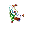
Mass: 18.015 Da / Num. of mol.: 92 / Source method: isolated from a natural source / Formula: H2O

-
Experimental details

-
Experiment

ExperimentMethod: X-RAY DIFFRACTION / Number of used crystals: 1

-
Sample preparation

CrystalDensity Matthews: 1.92 Å3/Da / Density % sol: 35.8 %
Crystal growTemperature: 293 K / Method: vapor diffusion, hanging drop / pH: 8.5 / Details: 0.1M Tris.HCl, 1.25M Ammonium sulphate

-
Data collection

DiffractionMean temperature: 100 K
Diffraction sourceSource: ROTATING ANODE / Type: RIGAKU MICROMAX-007 HF / Wavelength: 1.5418 Å
DetectorType: MAR scanner 345 mm plate / Detector: IMAGE PLATE / Date: Dec 20, 2012
RadiationMonochromator: multilayer mirrors Varimax HF / Protocol: SINGLE WAVELENGTH / Monochromatic (M) / Laue (L): M / Scattering type: x-ray
Radiation wavelengthWavelength: 1.5418 Å / Relative weight: 1
ReflectionResolution: 2.457→45.703 Å / Num. all: 8737 / Num. obs: 8737 / % possible obs: 98.9 % / Redundancy: 3.5 % / Biso Wilson estimate: 36.1 Å2 / Rpim(I) all: 0.067 / Rrim(I) all: 0.129 / Rsym value: 0.109 / Net I/av σ(I): 6.106 / Net I/σ(I): 10.2 / Num. measured all: 30830
Reflection shell

Diffraction-ID: 1 / Rejects: 0

Resolution (Å)Redundancy (%)Rmerge(I) obsMean I/σ(I) obsNum. measured allNum. unique allRpim(I) allRsym valueNet I/σ(I) obs% possible all
2.457-2.593.50.3782416511940.2320.3784.196.3
2.59-2.753.60.4471.7427811870.2720.4473.698.8
2.75-2.943.60.3242.4397711060.1990.3244.598.5
2.94-3.173.60.2043.8374610400.1240.2046.499.5
3.17-3.473.60.1097.134669680.0660.10910.699.6
3.47-3.883.60.06811.531798930.0410.06814.699.7
3.88-4.483.50.04517.228057970.0280.04518.299.9
4.48-5.493.50.04516.823686820.0280.04518.6100
5.49-7.773.40.04516.518485470.0280.04517.299.9
7.77-35.4213.10.02879983230.0210.02825.498.5

-
Processing

Software
NameVersionClassification
SCALA3.3.20data scaling
XDSdata scaling
PDB_EXTRACT3.14data extraction
BUSTER2.10.0refinement
RefinementMethod to determine structure: MOLECULAR REPLACEMENT
Starting model: a number of search probes were generated by comparative modelling with the Rosetta suite [Das, R. & Baker, D. Macromolecular modeling with rosetta (2008) Annu Rev Biochem 77, 363-82], ...Starting model: a number of search probes were generated by comparative modelling with the Rosetta suite [Das, R. & Baker, D. Macromolecular modeling with rosetta (2008) Annu Rev Biochem 77, 363-82], using PDB ID 3h47 as a template. In this way, 10000 models were obtained, scored and clustered. Several top ranking models gave solutions in MR using the program Phaser. To note, using PDB 3H47 (or any other available PDB with similar sequence) did not produce right solutions.
Resolution: 2.46→35.42 Å / Cor.coef. Fo:Fc: 0.9163 / Cor.coef. Fo:Fc free: 0.8752 / SU R Cruickshank DPI: 0.551 / Cross valid method: THROUGHOUT / σ(F): 0 / SU R Blow DPI: 0.725 / SU Rfree Blow DPI: 0.291 / SU Rfree Cruickshank DPI: 0.285
RfactorNum. reflection% reflectionSelection details
Rfree0.2533 867 9.96 %RANDOM
Rwork0.1993 ---
obs0.2047 8701 98.89 %-
Displacement parametersBiso max: 119.55 Å2 / Biso mean: 33.56 Å2 / Biso min: 8.55 Å2
Baniso -1Baniso -2Baniso -3
1-1.6214 Å20 Å20 Å2
2--5.3721 Å20 Å2
3----6.9935 Å2
Refine analyzeLuzzati coordinate error obs: 0.333 Å
Refinement stepCycle: final / Resolution: 2.46→35.42 Å
ProteinNucleic acidLigandSolventTotal
Num. atoms1723 0 0 92 1815
Biso mean---28.41 -
Num. residues----228
Refine LS restraints
Refine-IDTypeNumberRestraint functionWeightDev ideal
X-RAY DIFFRACTIONt_dihedral_angle_d572SINUSOIDAL2
X-RAY DIFFRACTIONt_trig_c_planes53HARMONIC2
X-RAY DIFFRACTIONt_gen_planes247HARMONIC5
X-RAY DIFFRACTIONt_it1761HARMONIC20
X-RAY DIFFRACTIONt_nbd
X-RAY DIFFRACTIONt_improper_torsion
X-RAY DIFFRACTIONt_pseud_angle
X-RAY DIFFRACTIONt_chiral_improper_torsion233SEMIHARMONIC5
X-RAY DIFFRACTIONt_sum_occupancies
X-RAY DIFFRACTIONt_utility_distance
X-RAY DIFFRACTIONt_utility_angle
X-RAY DIFFRACTIONt_utility_torsion
X-RAY DIFFRACTIONt_ideal_dist_contact2164SEMIHARMONIC4
X-RAY DIFFRACTIONt_bond_d1761HARMONIC20.01
X-RAY DIFFRACTIONt_angle_deg2407HARMONIC21.21
X-RAY DIFFRACTIONt_omega_torsion2.55
X-RAY DIFFRACTIONt_other_torsion20.23
LS refinement shellResolution: 2.46→2.75 Å / Total num. of bins used: 5
RfactorNum. reflection% reflection
Rfree0.3008 243 10.15 %
Rwork0.2193 2150 -
all0.2275 2393 -
obs--98.89 %
Refinement TLS params.

Method: refined / Refine-ID: X-RAY DIFFRACTION

IDL112)L122)L132)L222)L232)L332)S11 (Å °)S12 (Å °)S13 (Å °)S21 (Å °)S22 (Å °)S23 (Å °)S31 (Å °)S32 (Å °)S33 (Å °)T112)T122)T132)T222)T232)T332)Origin x (Å)Origin y (Å)Origin z (Å)
10.86641.2093-0.19155.01430.90871.46690.0531-0.0047-0.37550.14680.0082-0.0081-0.1245-0.0807-0.0613-0.0970.004-0.0118-0.1190.02210.0763-17.41698.25735.5747
23.0875-1.92250.17735.0865-0.59892.97-0.03880.00270.03120.17310.09910.1815-0.1702-0.0865-0.0603-0.0912-0.01930.0528-0.17040.04220.1203-20.5039-9.453312.6163
33.7693-0.38761.28934.2214-0.085.0855-0.00650.12730.087-0.29830.19160.0673-0.0975-0.3584-0.1851-0.15870.0044-0.0679-0.19590.01460.1783-39.3031.0699-7.4733
Refinement TLS group
IDRefine-IDRefine TLS-IDSelection detailsAuth asym-IDAuth seq-ID
1X-RAY DIFFRACTION1{ A|* }A129 - 206
2X-RAY DIFFRACTION2{ B|* }B131 - 208
3X-RAY DIFFRACTION3{ C|* }C129 - 207

+
About Yorodumi

-
News

-
Feb 9, 2022. New format data for meta-information of EMDB entries

New format data for meta-information of EMDB entries

  • Version 3 of the EMDB header file is now the official format.
  • The previous official version 1.9 will be removed from the archive.

Related info.:EMDB header

External links:wwPDB to switch to version 3 of the EMDB data model

-
Aug 12, 2020. Covid-19 info

Covid-19 info

URL: https://pdbj.org/emnavi/covid19.php

New page: Covid-19 featured information page in EM Navigator.

Related info.:Covid-19 info / Mar 5, 2020. Novel coronavirus structure data

+
Mar 5, 2020. Novel coronavirus structure data

Novel coronavirus structure data

Related info.:Yorodumi Speices / Aug 12, 2020. Covid-19 info

External links:COVID-19 featured content - PDBj / Molecule of the Month (242):Coronavirus Proteases

+
Jan 31, 2019. EMDB accession codes are about to change! (news from PDBe EMDB page)

EMDB accession codes are about to change! (news from PDBe EMDB page)

  • The allocation of 4 digits for EMDB accession codes will soon come to an end. Whilst these codes will remain in use, new EMDB accession codes will include an additional digit and will expand incrementally as the available range of codes is exhausted. The current 4-digit format prefixed with “EMD-” (i.e. EMD-XXXX) will advance to a 5-digit format (i.e. EMD-XXXXX), and so on. It is currently estimated that the 4-digit codes will be depleted around Spring 2019, at which point the 5-digit format will come into force.
  • The EM Navigator/Yorodumi systems omit the EMD- prefix.

Related info.:Q: What is EMD? / ID/Accession-code notation in Yorodumi/EM Navigator

External links:EMDB Accession Codes are Changing Soon! / Contact to PDBj

+
Jul 12, 2017. Major update of PDB

Major update of PDB

  • wwPDB released updated PDB data conforming to the new PDBx/mmCIF dictionary.
  • This is a major update changing the version number from 4 to 5, and with Remediation, in which all the entries are updated.
  • In this update, many items about electron microscopy experimental information are reorganized (e.g. em_software).
  • Now, EM Navigator and Yorodumi are based on the updated data.

External links:wwPDB Remediation / Enriched Model Files Conforming to OneDep Data Standards Now Available in the PDB FTP Archive

-
Yorodumi

Thousand views of thousand structures

  • Yorodumi is a browser for structure data from EMDB, PDB, SASBDB, etc.
  • This page is also the successor to EM Navigator detail page, and also detail information page/front-end page for Omokage search.
  • The word "yorodu" (or yorozu) is an old Japanese word meaning "ten thousand". "mi" (miru) is to see.

Related info.:EMDB / PDB / SASBDB / Comparison of 3 databanks / Yorodumi Search / Aug 31, 2016. New EM Navigator & Yorodumi / Yorodumi Papers / Jmol/JSmol / Function and homology information / Changes in new EM Navigator and Yorodumi

Read more