 Movie
Movie Controller

[English] 日本語
 Yorodumi
- PDB-4pf4: 1.1A X-RAY STRUCTURE OF THE APO CATALYTIC DOMAIN OF DEATH-ASSOCIA... -
+ Open data

- Basic information
| Entry | Database: PDB / ID: 4pf4 | ||||||
|---|---|---|---|---|---|---|---|
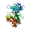
| Title | 1.1A X-RAY STRUCTURE OF THE APO CATALYTIC DOMAIN OF DEATH-ASSOCIATED PROTEIN KINASE 1, aa 1-277 | ||||||
 Components Components | Death-associated protein kinase 1 DAPK1 DAPK1 | ||||||
 Keywords Keywords | TRANSFERASE | ||||||
| Function / homology |  Function and homology information Function and homology informationcellular response to hydroperoxide / regulation of response to tumor cell / positive regulation of autophagic cell death / DAPK1-calmodulin complex / defense response to tumor cell / Caspase activation via Dependence Receptors in the absence of ligand / syntaxin-1 binding / regulation of NMDA receptor activity / calmodulin-dependent protein kinase activity / positive regulation of cysteine-type endopeptidase activity involved in apoptotic process ...cellular response to hydroperoxide / regulation of response to tumor cell / positive regulation of autophagic cell death / DAPK1-calmodulin complex / defense response to tumor cell / Caspase activation via Dependence Receptors in the absence of ligand / syntaxin-1 binding / regulation of NMDA receptor activity / calmodulin-dependent protein kinase activity / positive regulation of cysteine-type endopeptidase activity involved in apoptotic process / extrinsic apoptotic signaling pathway via death domain receptors / positive regulation of autophagy / regulation of autophagy / apoptotic signaling pathway / cellular response to type II interferon / actin cytoskeleton / regulation of apoptotic process / protein autophosphorylation / postsynaptic density / negative regulation of translation / calmodulin binding / non-specific serine/threonine protein kinase / protein kinase activity / intracellular signal transduction / positive regulation of apoptotic process / protein phosphorylation / protein serine kinase activity / protein serine/threonine kinase activity / glutamatergic synapse / apoptotic process / GTP binding / negative regulation of apoptotic process / ATP binding / identical protein binding / nucleus / plasma membrane / cytoplasmSimilarity search - Function | ||||||
| Biological species |  Homo sapiens (human) Homo sapiens (human) | ||||||
| Method | X-RAY DIFFRACTION / SYNCHROTRON / MOLECULAR REPLACEMENT / molecular replacement / Resolution: 1.13 Å | ||||||
 Authors Authors | Temmerman, K. / Simon, B. / Wilmanns, M. | ||||||
 Citation Citation | Journal: To Be Published Title: 1.1A X-RAY STRUCTURE OF THE APO CATALYTIC DOMAIN OF DEATH-ASSOCIATED PROTEIN KINASE 1, aa 1-277 Authors: Temmerman, K. / Simon, B. / Wilmanns, M. | ||||||
| History |
|
- Structure visualization
| Structure viewer | Molecule: MolmilJmol/JSmol |
|---|
- Downloads & links
-Download
| PDBx/mmCIF format | 4pf4.cif.gz | 267.1 KB | Display | PDBx/mmCIF format |
|---|---|---|---|---|
| PDB format | pdb4pf4.ent.gz | 214.8 KB | Display | PDB format |
| PDBx/mmJSON format | 4pf4.json.gz | Tree view | PDBx/mmJSON format | |
| Others |  Other downloads Other downloads |
-Validation report
| Arichive directory | https://data.pdbj.org/pub/pdb/validation_reports/pf/4pf4ftp://data.pdbj.org/pub/pdb/validation_reports/pf/4pf4 | HTTPS FTP |
|---|
-Related structure data
| Related structure data |  2w4jS S: Starting model for refinement |
|---|---|
| Similar structure data |


-Links
 PDBj
PDBj


- Assembly
| Deposited unit | 
| ||||||||
|---|---|---|---|---|---|---|---|---|---|
| 1 |
| ||||||||
| Unit cell |
| ||||||||
| Details | biological unit is the same as asym. |
-Components
-Protein , 1 types, 1 molecules A
| #1: Protein | DAPK1 / DAP kinase 1 Mass: 31990.469 Da / Num. of mol.: 1 Source method: isolated from a genetically manipulated source Source: (gene. exp.) Homo sapiens (human) / Gene: DAPK1, DAPK / Plasmid: pnEA-vHisGST / Production host:  Escherichia coli BL21 (bacteria) / Variant (production host): Rosetta2 Escherichia coli BL21 (bacteria) / Variant (production host): Rosetta2References: UniProt: P53355, non-specific serine/threonine protein kinase |
|---|
-Non-polymers , 5 types, 502 molecules 


| #2: Chemical | ChemComp-SO4 / Sulfate Mass: 96.063 Da / Num. of mol.: 4 / Source method: obtained synthetically / Formula: SO4 Mass: 96.063 Da / Num. of mol.: 4 / Source method: obtained synthetically / Formula: SO4#3: Chemical |  Mass: 22.990 Da / Num. of mol.: 2 / Source method: obtained synthetically / Formula: Na Mass: 22.990 Da / Num. of mol.: 2 / Source method: obtained synthetically / Formula: Na#4: Chemical | ChemComp-GOL / Glycerol Mass: 92.094 Da / Num. of mol.: 6 / Source method: obtained synthetically / Formula: C3H8O3 Mass: 92.094 Da / Num. of mol.: 6 / Source method: obtained synthetically / Formula: C3H8O3#5: Chemical | ChemComp-MG / |  Mass: 24.305 Da / Num. of mol.: 1 / Source method: obtained synthetically / Formula: Mg Mass: 24.305 Da / Num. of mol.: 1 / Source method: obtained synthetically / Formula: Mg#6: Water | ChemComp-HOH / | WaterMass: 18.015 Da / Num. of mol.: 489 / Source method: isolated from a natural source / Formula: H2O |
|---|
-Experimental details
-Experiment
| Experiment | Method: X-RAY DIFFRACTION / Number of used crystals: 1 |
|---|
- Sample preparation
| Crystal | Density Matthews: 1.99 Å3/Da / Density % sol: 38.25 % |
|---|---|
| Crystal grow | Temperature: 292 K / Method: vapor diffusion, sitting drop / pH: 7 / Details: 0.1 M HEPES, Ph=7, 2M Ammonium sulfate |
-Data collection
| Diffraction | Mean temperature: 100 K | ||||||||||||||||||||||||||||||||||||||||||||||||||||||||||||||||||||||||||||||||||||||||||||||||||||||||||||||
|---|---|---|---|---|---|---|---|---|---|---|---|---|---|---|---|---|---|---|---|---|---|---|---|---|---|---|---|---|---|---|---|---|---|---|---|---|---|---|---|---|---|---|---|---|---|---|---|---|---|---|---|---|---|---|---|---|---|---|---|---|---|---|---|---|---|---|---|---|---|---|---|---|---|---|---|---|---|---|---|---|---|---|---|---|---|---|---|---|---|---|---|---|---|---|---|---|---|---|---|---|---|---|---|---|---|---|---|---|---|---|---|
| Diffraction source | Source: SYNCHROTRON / Site: ESRF  / Beamline: ID23-1 / Wavelength: 0.973 Å / Beamline: ID23-1 / Wavelength: 0.973 Å | ||||||||||||||||||||||||||||||||||||||||||||||||||||||||||||||||||||||||||||||||||||||||||||||||||||||||||||||
| Detector | Type: DECTRIS PILATUS 6M-F / Detector: PIXEL / Date: Apr 6, 2014 | ||||||||||||||||||||||||||||||||||||||||||||||||||||||||||||||||||||||||||||||||||||||||||||||||||||||||||||||
| Radiation | Monochromator: Si(111) / Protocol: SINGLE WAVELENGTH / Monochromatic (M) / Laue (L): M / Scattering type: x-ray | ||||||||||||||||||||||||||||||||||||||||||||||||||||||||||||||||||||||||||||||||||||||||||||||||||||||||||||||
| Radiation wavelength | Wavelength: 0.973 Å / Relative weight: 1 | ||||||||||||||||||||||||||||||||||||||||||||||||||||||||||||||||||||||||||||||||||||||||||||||||||||||||||||||
| Reflection | Resolution: 1.129→50.758 Å / Num. all: 96009 / Num. obs: 96009 / % possible obs: 99.4 % / Redundancy: 6.3 % / Rpim(I) all: 0.032 / Rrim(I) all: 0.082 / Rsym value: 0.076 / Net I/av σ(I): 5.944 / Net I/σ(I): 10.7 / Num. measured all: 601419 | ||||||||||||||||||||||||||||||||||||||||||||||||||||||||||||||||||||||||||||||||||||||||||||||||||||||||||||||
| Reflection shell | Diffraction-ID: 1 / Rejects: 0
|
-Phasing
| Phasing | Method: molecular replacement | ||||||
|---|---|---|---|---|---|---|---|
| Phasing MR |
|
- Processing
| Software |
| |||||||||||||||||||||||||||||||||||||||||||||||||||||||||||||||||||||||||||
|---|---|---|---|---|---|---|---|---|---|---|---|---|---|---|---|---|---|---|---|---|---|---|---|---|---|---|---|---|---|---|---|---|---|---|---|---|---|---|---|---|---|---|---|---|---|---|---|---|---|---|---|---|---|---|---|---|---|---|---|---|---|---|---|---|---|---|---|---|---|---|---|---|---|---|---|---|
| Refinement | Method to determine structure: MOLECULAR REPLACEMENT Starting model: 2W4J Resolution: 1.13→50.758 Å / Cor.coef. Fo:Fc: 0.985 / Cor.coef. Fo:Fc free: 0.978 / WRfactor Rfree: 0.1467 / WRfactor Rwork: 0.1134 / FOM work R set: 0.9154 / SU B: 1.024 / SU ML: 0.021 / SU R Cruickshank DPI: 0.0284 / SU Rfree: 0.0307 / Cross valid method: THROUGHOUT / σ(F): 0 / ESU R: 0.028 / ESU R Free: 0.031 / Stereochemistry target values: MAXIMUM LIKELIHOOD Details: HYDROGENS HAVE BEEN ADDED IN THE RIDING POSITIONS U VALUES : REFINED INDIVIDUALLY
| |||||||||||||||||||||||||||||||||||||||||||||||||||||||||||||||||||||||||||
| Solvent computation | Ion probe radii: 0.8 Å / Shrinkage radii: 0.8 Å / VDW probe radii: 1.2 Å / Solvent model: MASK | |||||||||||||||||||||||||||||||||||||||||||||||||||||||||||||||||||||||||||
| Displacement parameters | Biso max: 144.22 Å2 / Biso mean: 16.198 Å2 / Biso min: 5.94 Å2
| |||||||||||||||||||||||||||||||||||||||||||||||||||||||||||||||||||||||||||
| Refinement step | Cycle: final / Resolution: 1.13→50.758 Å
| |||||||||||||||||||||||||||||||||||||||||||||||||||||||||||||||||||||||||||
| Refine LS restraints |
| |||||||||||||||||||||||||||||||||||||||||||||||||||||||||||||||||||||||||||
| LS refinement shell | Resolution: 1.129→1.158 Å / Total num. of bins used: 20
|
