 Movie
Movie Controller


+ Open data

- Basic information
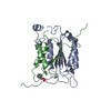
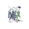
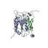
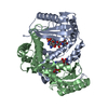
| Entry | Database: PDB / ID: 4pde | ||||||
|---|---|---|---|---|---|---|---|
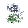
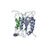
| Title | Crystal structure of FdhD in complex with GDP | ||||||
 Components Components | Protein FdhD | ||||||
 Keywords Keywords |  HYDROLASE / GDP / Mo-bisPGD sulfuration HYDROLASE / GDP / Mo-bisPGD sulfuration | ||||||
| Function / homology |  Function and homology information Function and homology informationsulfur carrier activity / sulfurtransferase activity / Mo-molybdopterin cofactor biosynthetic process / molybdopterin cofactor binding / positive regulation of catalytic activity / cytosol / cytoplasmSimilarity search - Function | ||||||
| Biological species |  Escherichia coli (E. coli) Escherichia coli (E. coli) | ||||||
| Method | X-RAY DIFFRACTION / SYNCHROTRON / MOLECULAR REPLACEMENT / Resolution: 2.8 Å | ||||||
 Authors Authors | Arnoux, P. / Walburger, A. / Magalon, A. / Pignol, D. | ||||||
 Citation Citation | Journal: Nat Commun / Year: 2015 Title: Sulphur shuttling across a chaperone during molybdenum cofactor maturation. Authors: Arnoux, P. / Ruppelt, C. / Oudouhou, F. / Lavergne, J. / Siponen, M.I. / Toci, R. / Mendel, R.R. / Bittner, F. / Pignol, D. / Magalon, A. / Walburger, A. | ||||||
| History |
|
- Structure visualization
| Structure viewer | Molecule: MolmilJmol/JSmol |
|---|
- Downloads & links
-Download
| PDBx/mmCIF format | 4pde.cif.gz | 111.7 KB | Display | PDBx/mmCIF format |
|---|---|---|---|---|
| PDB format | pdb4pde.ent.gz | 84.9 KB | Display | PDB format |
| PDBx/mmJSON format | 4pde.json.gz | Tree view | PDBx/mmJSON format | |
| Others |  Other downloads Other downloads |
-Validation report
| Arichive directory | https://data.pdbj.org/pub/pdb/validation_reports/pd/4pdeftp://data.pdbj.org/pub/pdb/validation_reports/pd/4pde | HTTPS FTP |
|---|
-Related structure data
| Related structure data |  1pw9S S: Starting model for refinement |
|---|---|
| Similar structure data |


-Links
 PDBj
PDBj- Assembly
| Deposited unit | 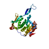
| ||||||||
|---|---|---|---|---|---|---|---|---|---|
| 1 | 
| ||||||||
| Unit cell |
|
-Components
| #1: Protein | Mass: 31668.137 Da / Num. of mol.: 1 Source method: isolated from a genetically manipulated source Source: (gene. exp.) Escherichia coli (E. coli) / Gene: fdhD, BN17_38461 / Production host: Escherichia coli (E. coli) / References: UniProt: J7QY30, UniProt: P32177*PLUS |
|---|---|
| #2: Chemical | ChemComp-GDP / Guanosine diphosphate  Type: RNA linking / Mass: 443.201 Da / Num. of mol.: 1 / Source method: obtained synthetically / Formula: C10H15N5O11P2 / Comment: GDP, energy-carrying molecule*YM Type: RNA linking / Mass: 443.201 Da / Num. of mol.: 1 / Source method: obtained synthetically / Formula: C10H15N5O11P2 / Comment: GDP, energy-carrying molecule*YM |
| #3: Water | ChemComp-HOH / Water Mass: 18.015 Da / Num. of mol.: 16 / Source method: isolated from a natural source / Formula: H2O Mass: 18.015 Da / Num. of mol.: 16 / Source method: isolated from a natural source / Formula: H2O |
-Experimental details
-Experiment
| Experiment | Method: X-RAY DIFFRACTION |
|---|
- Sample preparation
| Crystal | Density Matthews: 2.66 Å3/Da / Density % sol: 53.73 % |
|---|---|
| Crystal grow | Temperature: 293 K / Method: vapor diffusion, sitting drop / pH: 8 / Details: 0.2M NaNO3, 2.2 M AmSO4 |
-Data collection
| Diffraction | Mean temperature: 100 K |
|---|---|
| Diffraction source | Source: SYNCHROTRON / Site: ESRF  / Beamline: ID29 / Wavelength: 0.984 Å / Beamline: ID29 / Wavelength: 0.984 Å |
| Detector | Type: DECTRIS PILATUS 6M / Detector: PIXEL / Date: Jun 29, 2012 |
| Radiation | Protocol: SINGLE WAVELENGTH / Monochromatic (M) / Laue (L): M / Scattering type: x-ray |
| Radiation wavelength | Wavelength: 0.984 Å / Relative weight: 1 |
| Reflection | Resolution: 2.8→85 Å / Num. obs: 8948 / % possible obs: 99.8 % / Redundancy: 8.4 % / Net I/σ(I): 12.4 |
- Processing
| Software | Name: PHENIX / Version: (phenix.refine: 1.6_289) / Classification: refinement | |||||||||||||||||||||||||||||||||||||||||||||||||||||||||||||||||||||||||||||||||||||||||||||||||||||||||||||||||||||||||||||
|---|---|---|---|---|---|---|---|---|---|---|---|---|---|---|---|---|---|---|---|---|---|---|---|---|---|---|---|---|---|---|---|---|---|---|---|---|---|---|---|---|---|---|---|---|---|---|---|---|---|---|---|---|---|---|---|---|---|---|---|---|---|---|---|---|---|---|---|---|---|---|---|---|---|---|---|---|---|---|---|---|---|---|---|---|---|---|---|---|---|---|---|---|---|---|---|---|---|---|---|---|---|---|---|---|---|---|---|---|---|---|---|---|---|---|---|---|---|---|---|---|---|---|---|---|---|---|
| Refinement | Method to determine structure: MOLECULAR REPLACEMENT Starting model: 1pw9 Resolution: 2.8→69.573 Å / SU ML: 0.38 / Cross valid method: FREE R-VALUE / σ(F): 0.08 / Phase error: 25.59 / Stereochemistry target values: ML
| |||||||||||||||||||||||||||||||||||||||||||||||||||||||||||||||||||||||||||||||||||||||||||||||||||||||||||||||||||||||||||||
| Solvent computation | Shrinkage radii: 0.9 Å / VDW probe radii: 1.11 Å / Solvent model: FLAT BULK SOLVENT MODEL / Bsol: 39.814 Å2 / ksol: 0.369 e/Å3 | |||||||||||||||||||||||||||||||||||||||||||||||||||||||||||||||||||||||||||||||||||||||||||||||||||||||||||||||||||||||||||||
| Displacement parameters |
| |||||||||||||||||||||||||||||||||||||||||||||||||||||||||||||||||||||||||||||||||||||||||||||||||||||||||||||||||||||||||||||
| Refinement step | Cycle: LAST / Resolution: 2.8→69.573 Å
| |||||||||||||||||||||||||||||||||||||||||||||||||||||||||||||||||||||||||||||||||||||||||||||||||||||||||||||||||||||||||||||
| Refine LS restraints |
| |||||||||||||||||||||||||||||||||||||||||||||||||||||||||||||||||||||||||||||||||||||||||||||||||||||||||||||||||||||||||||||
| LS refinement shell |
| |||||||||||||||||||||||||||||||||||||||||||||||||||||||||||||||||||||||||||||||||||||||||||||||||||||||||||||||||||||||||||||
| Refinement TLS params. | Method: refined / Refine-ID: X-RAY DIFFRACTION
| |||||||||||||||||||||||||||||||||||||||||||||||||||||||||||||||||||||||||||||||||||||||||||||||||||||||||||||||||||||||||||||
| Refinement TLS group |
|
