 Movie
Movie Controller


+ Open data

- Basic information
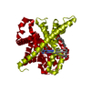
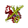
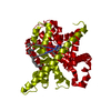
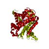
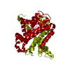
| Entry | Database: PDB / ID: 4pd5 | ||||||
|---|---|---|---|---|---|---|---|
| Title | Crystal structure of vcCNT-7C8C bound to gemcitabine | ||||||
 Components Components | NupC family protein | ||||||
 Keywords Keywords |  TRANSPORT PROTEIN / membrane protein / sodium-coupled transporter / gemcitabine / drug transporter TRANSPORT PROTEIN / membrane protein / sodium-coupled transporter / gemcitabine / drug transporter | ||||||
| Function / homology |  Function and homology information Function and homology informationnucleoside transmembrane transport / nucleoside transmembrane transporter activity / pyrimidine- and adenosine-specific:sodium symporter activity / purine-specific nucleoside:sodium symporter activity / pyrimidine nucleoside transport / purine nucleoside transmembrane transport / symporter activity / identical protein binding / plasma membraneSimilarity search - Function | ||||||
| Biological species |  Vibrio cholerae serotype O1 (bacteria) Vibrio cholerae serotype O1 (bacteria) | ||||||
| Method | X-RAY DIFFRACTION / SYNCHROTRON / MOLECULAR REPLACEMENT / Resolution: 2.906 Å | ||||||
 Authors Authors | Johnson, Z.L. / Lee, S.-Y. | ||||||
| Funding support |  United States, 1items United States, 1items
| ||||||
 Citation Citation | Journal: Elife / Year: 2014 Title: Structural basis of nucleoside and nucleoside drug selectivity by concentrative nucleoside transporters. Authors: Johnson, Z.L. / Lee, J.H. / Lee, K. / Lee, M. / Kwon, D.Y. / Hong, J. / Lee, S.Y. | ||||||
| History |
|
- Structure visualization
| Structure viewer | Molecule: MolmilJmol/JSmol |
|---|
- Downloads & links
-Download
| PDBx/mmCIF format | 4pd5.cif.gz | 90.7 KB | Display | PDBx/mmCIF format |
|---|---|---|---|---|
| PDB format | pdb4pd5.ent.gz | 65.2 KB | Display | PDB format |
| PDBx/mmJSON format | 4pd5.json.gz | Tree view | PDBx/mmJSON format | |
| Others |  Other downloads Other downloads |
-Validation report
| Arichive directory | https://data.pdbj.org/pub/pdb/validation_reports/pd/4pd5ftp://data.pdbj.org/pub/pdb/validation_reports/pd/4pd5 | HTTPS FTP |
|---|
-Related structure data
| Related structure data |  4pb1C  4pb2C  4pd6C  4pd7C  4pd8C  4pd9C  4pdaC  3tijS S: Starting model for refinement C: citing same article ( |
|---|---|
| Similar structure data |


-Links
 PDBj
PDBj- Assembly
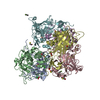
| Deposited unit | 
| ||||||||
|---|---|---|---|---|---|---|---|---|---|
| 1 | 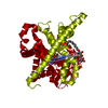
| ||||||||
| Unit cell |
| ||||||||
| Details | biological unit is the same as asym. |
-Components
| #1: Protein | Mass: 44151.848 Da / Num. of mol.: 1 / Mutation: L7C, I8C Source method: isolated from a genetically manipulated source Source: (gene. exp.) Vibrio cholerae serotype O1 (bacteria) / Strain: ATCC 39315 / El Tor Inaba N16961 / Gene: VC_2352 / Plasmid: pET26 / Production host: Escherichia coli (E. coli) / Strain (production host): C41 (DE3) / References: UniProt: Q9KPL5 |
|---|---|
| #2: Chemical | ChemComp-GEO / Gemcitabine  Mass: 263.198 Da / Num. of mol.: 1 / Source method: obtained synthetically / Formula: C9H11F2N3O4 / Comment: medication, chemotherapy*YM Mass: 263.198 Da / Num. of mol.: 1 / Source method: obtained synthetically / Formula: C9H11F2N3O4 / Comment: medication, chemotherapy*YM |
| #3: Chemical | ChemComp-NA /   Mass: 22.990 Da / Num. of mol.: 1 / Source method: obtained synthetically / Formula: Na Mass: 22.990 Da / Num. of mol.: 1 / Source method: obtained synthetically / Formula: Na |
| #4: Sugar | ChemComp-DMU /   Type: D-saccharide / Mass: 482.562 Da / Num. of mol.: 1 Type: D-saccharide / Mass: 482.562 Da / Num. of mol.: 1Source method: isolated from a genetically manipulated source Formula: C22H42O11 / Comment: detergent*YM |
| #5: Water | ChemComp-HOH / Water Mass: 18.015 Da / Num. of mol.: 9 / Source method: isolated from a natural source / Formula: H2O Mass: 18.015 Da / Num. of mol.: 9 / Source method: isolated from a natural source / Formula: H2O |
-Experimental details
-Experiment
| Experiment | Method: X-RAY DIFFRACTION / Number of used crystals: 1 |
|---|
- Sample preparation
| Crystal | Density Matthews: 4.01 Å3/Da / Density % sol: 69.29 % |
|---|---|
| Crystal grow | Temperature: 290 K / Method: liquid diffusion / pH: 8.5 / Details: 37-42% PEG400, 100 mM CaCl2 |
-Data collection
| Diffraction | Mean temperature: 100 K | ||||||||||||||||||||||||||||||||||||||||||||||||||||||||||||||||||||||||||||||||||||||||||||||||
|---|---|---|---|---|---|---|---|---|---|---|---|---|---|---|---|---|---|---|---|---|---|---|---|---|---|---|---|---|---|---|---|---|---|---|---|---|---|---|---|---|---|---|---|---|---|---|---|---|---|---|---|---|---|---|---|---|---|---|---|---|---|---|---|---|---|---|---|---|---|---|---|---|---|---|---|---|---|---|---|---|---|---|---|---|---|---|---|---|---|---|---|---|---|---|---|---|---|
| Diffraction source | Source: SYNCHROTRON / Site: APS / Beamline: 24-ID-C / Wavelength: 1.0004 Å | ||||||||||||||||||||||||||||||||||||||||||||||||||||||||||||||||||||||||||||||||||||||||||||||||
| Detector | Type: ADSC QUANTUM 315 / Detector: CCD / Date: Apr 9, 2012 | ||||||||||||||||||||||||||||||||||||||||||||||||||||||||||||||||||||||||||||||||||||||||||||||||
| Radiation | Protocol: SINGLE WAVELENGTH / Monochromatic (M) / Laue (L): M / Scattering type: x-ray | ||||||||||||||||||||||||||||||||||||||||||||||||||||||||||||||||||||||||||||||||||||||||||||||||
| Radiation wavelength | Wavelength: 1.0004 Å / Relative weight: 1 | ||||||||||||||||||||||||||||||||||||||||||||||||||||||||||||||||||||||||||||||||||||||||||||||||
| Reflection | Resolution: 2.9→50 Å / Num. obs: 14694 / % possible obs: 99.6 % / Redundancy: 5.7 % / Biso Wilson estimate: 82.67 Å2 / Rmerge(I) obs: 0.061 / Χ2: 1.059 / Net I/av σ(I): 30.765 / Net I/σ(I): 11.5 / Num. measured all: 83148 | ||||||||||||||||||||||||||||||||||||||||||||||||||||||||||||||||||||||||||||||||||||||||||||||||
| Reflection shell | Diffraction-ID: 1 / Rejects: 0
|
- Processing
| Software |
| ||||||||||||||||||||||||||||||||||||||||||
|---|---|---|---|---|---|---|---|---|---|---|---|---|---|---|---|---|---|---|---|---|---|---|---|---|---|---|---|---|---|---|---|---|---|---|---|---|---|---|---|---|---|---|---|
| Refinement | Method to determine structure: MOLECULAR REPLACEMENT Starting model: 3TIJ Resolution: 2.906→43.684 Å / FOM work R set: 0.7644 / SU ML: 0.38 / Cross valid method: FREE R-VALUE / σ(F): 1.39 / Phase error: 30.34 / Stereochemistry target values: ML
| ||||||||||||||||||||||||||||||||||||||||||
| Solvent computation | Shrinkage radii: 0.9 Å / VDW probe radii: 1.11 Å / Solvent model: FLAT BULK SOLVENT MODEL | ||||||||||||||||||||||||||||||||||||||||||
| Displacement parameters | Biso max: 186.47 Å2 / Biso mean: 76.81 Å2 / Biso min: 38.93 Å2 | ||||||||||||||||||||||||||||||||||||||||||
| Refinement step | Cycle: final / Resolution: 2.906→43.684 Å
| ||||||||||||||||||||||||||||||||||||||||||
| Refine LS restraints |
| ||||||||||||||||||||||||||||||||||||||||||
| LS refinement shell | Refine-ID: X-RAY DIFFRACTION / Total num. of bins used: 5
|
