 Movie
Movie Controller

[English] 日本語
 Yorodumi
- PDB-4p1c: CRYSTAL STRUCTURE OF THE TOLUENE 4-MONOOXYGENASE HYDROXYLASE-FERR... -
+ Open data

- Basic information
| Entry | Database: PDB / ID: 4p1c | |||||||||
|---|---|---|---|---|---|---|---|---|---|---|
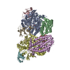
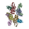
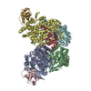
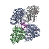
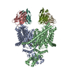
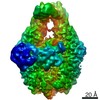
| Title | CRYSTAL STRUCTURE OF THE TOLUENE 4-MONOOXYGENASE HYDROXYLASE-FERREDOXIN C7S, C84A, C85A VARIANT ELECTRON-TRANSFER COMPLEX | |||||||||
 Components Components |
| |||||||||
 Keywords Keywords |  OXIDOREDUCTASE / ELECTRON-TRANSFER COMPLEX / DIIRON ENZYME COMPLEX / IRON-SULFUR / REDUCTION / HYDROXYLASE FERREDOXIN / OXYGENASE OXIDOREDUCTASE / ELECTRON-TRANSFER COMPLEX / DIIRON ENZYME COMPLEX / IRON-SULFUR / REDUCTION / HYDROXYLASE FERREDOXIN / OXYGENASE | |||||||||
| Function / homology |  Function and homology information Function and homology informationtoluene 4-monooxygenase / toluene catabolic process / oxidoreductase activity, acting on paired donors, with incorporation or reduction of molecular oxygen, NAD(P)H as one donor, and incorporation of one atom of oxygen / monooxygenase activity / 2 iron, 2 sulfur cluster binding / metal ion bindingSimilarity search - Function | |||||||||
| Biological species |  Pseudomonas mendocina (bacteria) Pseudomonas mendocina (bacteria) | |||||||||
| Method | X-RAY DIFFRACTION / SYNCHROTRON / MOLECULAR REPLACEMENT / Resolution: 2.4 Å | |||||||||
 Authors Authors | Acheson, J.F. / Fox, B.G. | |||||||||
| Funding support |  United States, 2items United States, 2items
| |||||||||
 Citation Citation | Journal: Nat Commun / Year: 2014 Title: Structural basis for biomolecular recognition in overlapping binding sites in a diiron enzyme system. Authors: Acheson, J.F. / Bailey, L.J. / Elsen, N.L. / Fox, B.G. | |||||||||
| History |
|
- Structure visualization
| Structure viewer | Molecule: MolmilJmol/JSmol |
|---|
- Downloads & links
-Download
| PDBx/mmCIF format | 4p1c.cif.gz | 419.4 KB | Display | PDBx/mmCIF format |
|---|---|---|---|---|
| PDB format | pdb4p1c.ent.gz | 337.8 KB | Display | PDB format |
| PDBx/mmJSON format | 4p1c.json.gz | Tree view | PDBx/mmJSON format | |
| Others |  Other downloads Other downloads |
-Validation report
| Arichive directory | https://data.pdbj.org/pub/pdb/validation_reports/p1/4p1cftp://data.pdbj.org/pub/pdb/validation_reports/p1/4p1c | HTTPS FTP |
|---|
-Related structure data
| Related structure data |  4p1bC  3dhgS S: Starting model for refinement C: citing same article ( |
|---|---|
| Similar structure data |

-Links
 PDBj
PDBj


- Assembly
| Deposited unit | 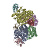
| ||||||||
|---|---|---|---|---|---|---|---|---|---|
| 1 |
| ||||||||
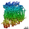
| Unit cell |
|
-Components
-Toluene-4-monooxygenase system protein ... , 3 types, 6 molecules ADBECF
| #1: Protein | Mass: 57089.910 Da / Num. of mol.: 2 / Mutation: Stop>492 Source method: isolated from a genetically manipulated source Source: (gene. exp.) Pseudomonas mendocina (bacteria) / Gene: tmoA / Plasmid: PVP58KABE3 / Production host: Escherichia coli (E. coli) / Strain (production host): BL21References: UniProt: Q00456, Oxidoreductases; Acting on paired donors, with incorporation or reduction of molecular oxygen; With NADH or NADPH as one donor, and incorporation of one atom of oxygen into the other donor#2: Protein | Mass: 35930.363 Da / Num. of mol.: 2 / Mutation: STOP>307 Source method: isolated from a genetically manipulated source Source: (gene. exp.) Pseudomonas mendocina (bacteria) / Gene: tmoE / Plasmid: PVP58KABE3 / Production host: Escherichia coli (E. coli) / Strain (production host): BL21References: UniProt: Q00460, Oxidoreductases; Acting on paired donors, with incorporation or reduction of molecular oxygen; With NADH or NADPH as one donor, and incorporation of one atom of oxygen into the other donor#3: Protein | Mass: 9340.679 Da / Num. of mol.: 2 Source method: isolated from a genetically manipulated source Source: (gene. exp.) Pseudomonas mendocina (bacteria) / Gene: tmoB / Plasmid: PVP58KABE3 / Production host: Escherichia coli (E. coli) / Strain (production host): BL21References: UniProt: Q00457, Oxidoreductases; Acting on paired donors, with incorporation or reduction of molecular oxygen; With NADH or NADPH as one donor, and incorporation of one atom of oxygen into the other donor |
|---|
-Protein , 1 types, 2 molecules HI
| #4: Protein | Mass: 12125.305 Da / Num. of mol.: 2 / Mutation: C7S C84A C85A Source method: isolated from a genetically manipulated source Source: (gene. exp.) Pseudomonas mendocina (bacteria) / Gene: tmoC / Plasmid: PET15BCDTET / Details (production host): C7S C84A C85A / Production host: Escherichia coli (E. coli) / Strain (production host): BL21 / References: UniProt: Q00458 |
|---|
-Non-polymers , 4 types, 617 molecules 


| #5: Chemical | ChemComp-FE / Iron Mass: 55.845 Da / Num. of mol.: 4 / Source method: obtained synthetically / Formula: Fe Mass: 55.845 Da / Num. of mol.: 4 / Source method: obtained synthetically / Formula: Fe#6: Chemical | Diethylene glycol Mass: 106.120 Da / Num. of mol.: 2 / Source method: obtained synthetically / Formula: C4H10O3 Mass: 106.120 Da / Num. of mol.: 2 / Source method: obtained synthetically / Formula: C4H10O3#7: Chemical | Iron–sulfur cluster Mass: 175.820 Da / Num. of mol.: 2 / Source method: obtained synthetically / Formula: Fe2S2 Mass: 175.820 Da / Num. of mol.: 2 / Source method: obtained synthetically / Formula: Fe2S2#8: Water | ChemComp-HOH / | WaterMass: 18.015 Da / Num. of mol.: 609 / Source method: isolated from a natural source / Formula: H2O |
|---|
-Details
| Sequence details | W336 AND Y227 ARE THE RESIDUES IN STRUCTURE. THERE MAY BE ERRORS IN THE ORIGINAL SEQUENCING OF THE ...W336 AND Y227 ARE THE RESIDUES IN STRUCTURE. THERE MAY BE ERRORS IN THE ORIGINAL SEQUENCING |
|---|
-Experimental details
-Experiment
| Experiment | Method: X-RAY DIFFRACTION / Number of used crystals: 1 |
|---|
- Sample preparation
| Crystal | Density Matthews: 2.36 Å3/Da / Density % sol: 47.88 % |
|---|---|
| Crystal grow | Temperature: 292 K / Method: vapor diffusion, hanging drop / pH: 7.5 Details: 100 MM MOPS/HEPES, 20% PEG 3350, 5% JEFFAMINE 200 MM AMMONIUM CHLORIDE, 10 MM MGCL2, PH 7.5, VAPOR DIFFUSION, TEMPERATURE 292K |
-Data collection
| Diffraction | Mean temperature: 100 K |
|---|---|
| Diffraction source | Source: SYNCHROTRON / Site: APS / Beamline: 21-ID-G / Wavelength: 0.97857 Å |
| Detector | Type: MARMOSAIC 300 mm CCD / Detector: CCD / Date: Nov 22, 2011 |
| Radiation | Monochromator: C(111) / Protocol: SINGLE WAVELENGTH / Monochromatic (M) / Laue (L): M / Scattering type: x-ray |
| Radiation wavelength | Wavelength: 0.97857 Å / Relative weight: 1 |
| Reflection | Resolution: 2.4→47.69 Å / Num. obs: 81284 / % possible obs: 94.6 % / Observed criterion σ(I): 1.7 / Redundancy: 6 % / Biso Wilson estimate: 27.96 Å2 / Rsym value: 0.129 / Net I/σ(I): 12.4 |
| Reflection shell | Resolution: 2.4→2.43 Å / Redundancy: 4.5 % / Mean I/σ(I) obs: 2 / Rsym value: 0.76 / % possible all: 93 |
- Processing
| Software |
| ||||||||||||||||||||||||||||||||||||||||||||||||||||||||||||||||||||||||||||||||||||||||||||||||||||||||||||||||||||||||||||||||||||||||||||||||||||||||||||||||||||||||||||||||||||||||||||||||||||||||||||||||||
|---|---|---|---|---|---|---|---|---|---|---|---|---|---|---|---|---|---|---|---|---|---|---|---|---|---|---|---|---|---|---|---|---|---|---|---|---|---|---|---|---|---|---|---|---|---|---|---|---|---|---|---|---|---|---|---|---|---|---|---|---|---|---|---|---|---|---|---|---|---|---|---|---|---|---|---|---|---|---|---|---|---|---|---|---|---|---|---|---|---|---|---|---|---|---|---|---|---|---|---|---|---|---|---|---|---|---|---|---|---|---|---|---|---|---|---|---|---|---|---|---|---|---|---|---|---|---|---|---|---|---|---|---|---|---|---|---|---|---|---|---|---|---|---|---|---|---|---|---|---|---|---|---|---|---|---|---|---|---|---|---|---|---|---|---|---|---|---|---|---|---|---|---|---|---|---|---|---|---|---|---|---|---|---|---|---|---|---|---|---|---|---|---|---|---|---|---|---|---|---|---|---|---|---|---|---|---|---|---|---|---|---|
| Refinement | Method to determine structure: MOLECULAR REPLACEMENT Starting model: 3DHG Resolution: 2.4→47.69 Å / SU ML: 0.27 / Cross valid method: FREE R-VALUE / σ(F): 1.34 / Phase error: 21.24 / Stereochemistry target values: ML
| ||||||||||||||||||||||||||||||||||||||||||||||||||||||||||||||||||||||||||||||||||||||||||||||||||||||||||||||||||||||||||||||||||||||||||||||||||||||||||||||||||||||||||||||||||||||||||||||||||||||||||||||||||
| Solvent computation | Shrinkage radii: 0.9 Å / VDW probe radii: 1.11 Å / Solvent model: FLAT BULK SOLVENT MODEL | ||||||||||||||||||||||||||||||||||||||||||||||||||||||||||||||||||||||||||||||||||||||||||||||||||||||||||||||||||||||||||||||||||||||||||||||||||||||||||||||||||||||||||||||||||||||||||||||||||||||||||||||||||
| Displacement parameters | Biso mean: 20.16 Å2 | ||||||||||||||||||||||||||||||||||||||||||||||||||||||||||||||||||||||||||||||||||||||||||||||||||||||||||||||||||||||||||||||||||||||||||||||||||||||||||||||||||||||||||||||||||||||||||||||||||||||||||||||||||
| Refinement step | Cycle: LAST / Resolution: 2.4→47.69 Å
| ||||||||||||||||||||||||||||||||||||||||||||||||||||||||||||||||||||||||||||||||||||||||||||||||||||||||||||||||||||||||||||||||||||||||||||||||||||||||||||||||||||||||||||||||||||||||||||||||||||||||||||||||||
| Refine LS restraints |
| ||||||||||||||||||||||||||||||||||||||||||||||||||||||||||||||||||||||||||||||||||||||||||||||||||||||||||||||||||||||||||||||||||||||||||||||||||||||||||||||||||||||||||||||||||||||||||||||||||||||||||||||||||
| LS refinement shell |
|
