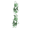
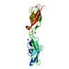
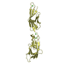
Entry Database : PDB / ID : 4oy9Title Crystal structure of human P-Cadherin EC1-EC2 in closed conformation Cadherin-3 Keywords / / / Function / homology Biological species Homo sapiens (human)Method / / Resolution : 1.62 Å Authors Dalle Vedove, A. / Lucarelli, A.P. / Nardone, V. / Matino, A. / Parisini, E. Funding support European Union, 1items Organization Grant number Country European Union (EU) FP7-PEOPLE-2010-RG (N. 268231) European Union
Journal : Acta Crystallogr.,Sect.F / Year : 2015Title : The X-ray structure of human P-cadherin EC1-EC2 in a closed conformation provides insight into the type I cadherin dimerization pathway.Authors : Dalle Vedove, A. / Lucarelli, A.P. / Nardone, V. / Matino, A. / Parisini, E. History Deposition Feb 11, 2014 Deposition site / Processing site Revision 1.0 Apr 1, 2015 Provider / Type Revision 1.1 Apr 22, 2015 Group Revision 1.2 Jan 8, 2020 Group Author supporting evidence / Derived calculations ... Author supporting evidence / Derived calculations / Other / Source and taxonomy / Structure summary Category entity_src_gen / pdbx_audit_support ... entity_src_gen / pdbx_audit_support / pdbx_database_status / pdbx_struct_assembly / pdbx_struct_conn_angle / pdbx_struct_oper_list / struct_keywords Item _entity_src_gen.pdbx_alt_source_flag / _pdbx_audit_support.country ... _entity_src_gen.pdbx_alt_source_flag / _pdbx_audit_support.country / _pdbx_audit_support.funding_organization / _pdbx_database_status.pdb_format_compatible / _pdbx_struct_assembly.oligomeric_details / _pdbx_struct_oper_list.symmetry_operation / _struct_keywords.text Revision 1.3 Dec 27, 2023 Group Data collection / Database references ... Data collection / Database references / Derived calculations / Refinement description Category chem_comp_atom / chem_comp_bond ... chem_comp_atom / chem_comp_bond / database_2 / pdbx_struct_conn_angle / refine_hist / struct_conn Item _database_2.pdbx_DOI / _database_2.pdbx_database_accession ... _database_2.pdbx_DOI / _database_2.pdbx_database_accession / _pdbx_struct_conn_angle.ptnr1_auth_comp_id / _pdbx_struct_conn_angle.ptnr1_auth_seq_id / _pdbx_struct_conn_angle.ptnr1_label_asym_id / _pdbx_struct_conn_angle.ptnr1_label_atom_id / _pdbx_struct_conn_angle.ptnr1_label_comp_id / _pdbx_struct_conn_angle.ptnr1_label_seq_id / _pdbx_struct_conn_angle.ptnr2_auth_seq_id / _pdbx_struct_conn_angle.ptnr2_label_asym_id / _pdbx_struct_conn_angle.ptnr3_auth_comp_id / _pdbx_struct_conn_angle.ptnr3_auth_seq_id / _pdbx_struct_conn_angle.ptnr3_label_asym_id / _pdbx_struct_conn_angle.ptnr3_label_atom_id / _pdbx_struct_conn_angle.ptnr3_label_comp_id / _pdbx_struct_conn_angle.ptnr3_label_seq_id / _pdbx_struct_conn_angle.value / _refine_hist.number_atoms_solvent / _refine_hist.number_atoms_total / _refine_hist.pdbx_number_atoms_ligand / _refine_hist.pdbx_number_atoms_nucleic_acid / _refine_hist.pdbx_number_atoms_protein / _struct_conn.pdbx_dist_value / _struct_conn.ptnr1_label_atom_id / _struct_conn.ptnr2_auth_seq_id / _struct_conn.ptnr2_label_asym_id
Show all Show less
 Movie
Movie Controller

 Yorodumi
 Open data
 Basic information
 Components
 Keywords
 Function and homology information
 Authors
 Citation
 Structure visualization
 Downloads & links
 Other downloads
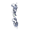

 PDBj
PDBj

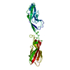
 Assembly


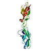
 Mass: 40.078 Da / Num. of mol.: 3 / Source method: obtained synthetically / Formula: Ca
 Mass: 18.015 Da / Num. of mol.: 251 / Source method: isolated from a natural source / Formula: H2O
 Sample preparation
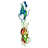
 / Beamline: X06DA / Wavelength: 1 Å
 Processing