| Entry | Database: PDB / ID: 4oxm
|
|---|
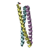
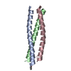
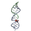
| Title | CRYSTAL STRUCTURE OF Central Coiled-Coil from Influenza Hemagglutinin HA2 without Heptad Repeat Stutter |
|---|
 Components Components | HA2-Del |
|---|
 Keywords Keywords |  VIRAL PROTEIN / coiled-coil / Influenza / Hemagglutinin / Stutter VIRAL PROTEIN / coiled-coil / Influenza / Hemagglutinin / Stutter |
|---|
| Function / homology | Haemagglutinin, influenzavirus B / Haemagglutinin / Haemagglutinin, influenzavirus A/B / host cell surface receptor binding / fusion of virus membrane with host plasma membrane / viral envelope / Truncated hemagglutinin Function and homology information Function and homology information |
|---|
| Biological species |   unidentified influenza virus unidentified influenza virus |
|---|
| Method | X-RAY DIFFRACTION / SYNCHROTRON / MOLECULAR REPLACEMENT / molecular replacement / Resolution: 1.9 Å |
|---|
 Authors Authors | Malashkevich, V.N. / Higgins, C.D. / Lai, J.R. / Almo, S.C. |
|---|
 Citation Citation | Journal: to be published
Title: CRYSTAL STRUCTURE OF Central Coiled-Coil from InfluenzaHemagglutinin HA2 without Heptad Repeat Stutter
Authors: Malashkevich, V.N. / Higgins, C.D. / Lai, J.R. / Almo, S.C. |
|---|
| History | | Deposition | Feb 5, 2014 | Deposition site: RCSB / Processing site: RCSB |
|---|
| Revision 1.0 | Apr 30, 2014 | Provider: repository / Type: Initial release |
|---|
| Revision 1.1 | Dec 24, 2014 | Group: Database references |
|---|
| Revision 1.2 | Nov 22, 2017 | Group: Database references / Derived calculations ...Database references / Derived calculations / Other / Refinement description / Source and taxonomy
Category: citation / pdbx_database_status ...citation / pdbx_database_status / pdbx_entity_src_syn / pdbx_struct_assembly / pdbx_struct_oper_list / software
Item: _citation.journal_id_CSD / _pdbx_database_status.pdb_format_compatible ..._citation.journal_id_CSD / _pdbx_database_status.pdb_format_compatible / _pdbx_entity_src_syn.pdbx_alt_source_flag / _pdbx_struct_assembly.oligomeric_details / _pdbx_struct_oper_list.symmetry_operation |
|---|
| Revision 1.3 | Dec 27, 2023 | Group: Data collection / Database references / Refinement description
Category: chem_comp_atom / chem_comp_bond ...chem_comp_atom / chem_comp_bond / database_2 / refine_hist / struct_ncs_dom_lim
Item: _database_2.pdbx_DOI / _database_2.pdbx_database_accession ..._database_2.pdbx_DOI / _database_2.pdbx_database_accession / _refine_hist.d_res_low / _refine_hist.number_atoms_solvent / _refine_hist.number_atoms_total / _refine_hist.pdbx_number_atoms_ligand / _refine_hist.pdbx_number_atoms_nucleic_acid / _refine_hist.pdbx_number_atoms_protein / _struct_ncs_dom_lim.beg_auth_comp_id / _struct_ncs_dom_lim.beg_label_asym_id / _struct_ncs_dom_lim.beg_label_comp_id / _struct_ncs_dom_lim.beg_label_seq_id / _struct_ncs_dom_lim.end_auth_comp_id / _struct_ncs_dom_lim.end_label_asym_id / _struct_ncs_dom_lim.end_label_comp_id / _struct_ncs_dom_lim.end_label_seq_id |
|---|
| Revision 1.4 | Mar 27, 2024 | Group: Refinement description / Category: pdbx_initial_refinement_model |
|---|
|
|---|
 Movie
Movie Controller

 Yorodumi
 Open data
 Basic information
 Structure visualization
 Downloads & links
 Other downloads


 PDBj
PDBj


 Assembly
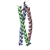
 Mass: 18.015 Da / Num. of mol.: 70 / Source method: isolated from a natural source / Formula: H2O
 Sample preparation
 / Beamline: 31-ID / Wavelength: 0.9791 Å
 Processing