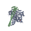
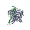
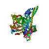
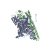
[English] 日本語
Yorodumi
- PDB-4ovu: Crystal Structure of p110alpha in complex with niSH2 of p85alpha -

+
Open data


ID or keywords:

Loading...

-
Basic information

Entry
Database: PDB / ID: 4ovu
TitleCrystal Structure of p110alpha in complex with niSH2 of p85alpha
Components
  • Phosphatidylinositol 3-kinase regulatory subunit alpha
  • Phosphatidylinositol 4,5-bisphosphate 3-kinase catalytic subunit alpha isoform
KeywordsTRANSFERASE/TRANSFERASE REGULATOR / p110 / p85 / PI3KCA / PI3K / PIK3R1 / phosphatidilynositol 3 / 4 / 5 triphosphate / PIP2 / phosphatidylinositol 4 / 5 bisphosphate / lipid kinase / phosphoinositide / 3-kinase / signaling / phosphatidylinositol 3-kinase / TRANSFERASE-TRANSFERASE REGULATOR complex
Function / homology
Function and homology information


perinuclear endoplasmic reticulum membrane / response to muscle inactivity / negative regulation of actin filament depolymerization / regulation of toll-like receptor 4 signaling pathway / response to L-leucine / phosphatidylinositol kinase activity / regulation of actin filament organization / phosphatidylinositol 3-kinase regulator activity / response to butyrate / autosome genomic imprinting ...perinuclear endoplasmic reticulum membrane / response to muscle inactivity / negative regulation of actin filament depolymerization / regulation of toll-like receptor 4 signaling pathway / response to L-leucine / phosphatidylinositol kinase activity / regulation of actin filament organization / phosphatidylinositol 3-kinase regulator activity / response to butyrate / autosome genomic imprinting / positive regulation of focal adhesion disassembly / IRS-mediated signalling / cellular response to hydrostatic pressure / phosphatidylinositol 3-kinase activator activity / interleukin-18-mediated signaling pathway / PI3K events in ERBB4 signaling / myeloid leukocyte migration / phosphatidylinositol 3-kinase regulatory subunit binding / 1-phosphatidylinositol-3-kinase regulator activity / neurotrophin TRKA receptor binding / Activated NTRK2 signals through PI3K / positive regulation of protein localization to membrane / cis-Golgi network / Activated NTRK3 signals through PI3K / kinase activator activity / ErbB-3 class receptor binding / negative regulation of fibroblast apoptotic process / RHOF GTPase cycle / cardiac muscle cell contraction / phosphatidylinositol 3-kinase complex, class IB / vasculature development / transmembrane receptor protein tyrosine kinase adaptor activity / RHOD GTPase cycle / positive regulation of endoplasmic reticulum unfolded protein response / Signaling by cytosolic FGFR1 fusion mutants / regulation of cellular respiration / enzyme-substrate adaptor activity / phosphatidylinositol 3-kinase complex / anoikis / Nephrin family interactions / 1-phosphatidylinositol-4-phosphate 3-kinase activity / RND1 GTPase cycle / Costimulation by the CD28 family / vascular endothelial growth factor signaling pathway / 1-phosphatidylinositol-4,5-bisphosphate 3-kinase activity / RND2 GTPase cycle / MET activates PI3K/AKT signaling / PI3K/AKT activation / RND3 GTPase cycle / positive regulation of leukocyte migration / phosphatidylinositol-4,5-bisphosphate 3-kinase / positive regulation of filopodium assembly / phosphatidylinositol 3-kinase complex, class IA / negative regulation of stress fiber assembly / phosphatidylinositol 3-kinase / relaxation of cardiac muscle / growth hormone receptor signaling pathway / phosphatidylinositol-3-phosphate biosynthetic process / insulin binding / 1-phosphatidylinositol-3-kinase activity / RHOV GTPase cycle / negative regulation of macroautophagy / RHOB GTPase cycle / Signaling by ALK / negative regulation of cell-matrix adhesion / GP1b-IX-V activation signalling / PI-3K cascade:FGFR3 / Erythropoietin activates Phosphoinositide-3-kinase (PI3K) / protein kinase activator activity / response to dexamethasone / PI-3K cascade:FGFR2 / PI-3K cascade:FGFR4 / RHOJ GTPase cycle / RHOC GTPase cycle / PI-3K cascade:FGFR1 / negative regulation of osteoclast differentiation / intracellular glucose homeostasis / CD28 dependent PI3K/Akt signaling / Synthesis of PIPs at the plasma membrane / phosphatidylinositol phosphate biosynthetic process / CDC42 GTPase cycle / RHOU GTPase cycle / PI3K events in ERBB2 signaling / Signaling by ALK fusions and activated point mutants / RHOG GTPase cycle / negative regulation of anoikis / RET signaling / T cell differentiation / regulation of multicellular organism growth / extrinsic apoptotic signaling pathway via death domain receptors / insulin receptor substrate binding / Interleukin-3, Interleukin-5 and GM-CSF signaling / PI3K Cascade / intercalated disc / RHOA GTPase cycle / positive regulation of TOR signaling / endothelial cell migration / RAC2 GTPase cycle / RAC3 GTPase cycle / GAB1 signalosome
Similarity search - Function
Helix Hairpins - #1490 / PI3Kalpha, catalytic domain / Phosphatidylinositol 3-kinase regulatory subunit alpha, SH3 domain / PIK3R1, inter-SH2 domain / PI3K p85 subunit, C-terminal SH2 domain / PI3K regulatory subunit p85-related , inter-SH2 domain / PI3K p85 subunit, N-terminal SH2 domain / Phosphatidylinositol 3-kinase regulatory subunit P85 inter-SH2 domain / Phosphatidylinositol 3-/4-kinase, catalytic domain / Phosphatidylinositol 3-kinase, accessory domain (PIK) ...Helix Hairpins - #1490 / PI3Kalpha, catalytic domain / Phosphatidylinositol 3-kinase regulatory subunit alpha, SH3 domain / PIK3R1, inter-SH2 domain / PI3K p85 subunit, C-terminal SH2 domain / PI3K regulatory subunit p85-related , inter-SH2 domain / PI3K p85 subunit, N-terminal SH2 domain / Phosphatidylinositol 3-kinase regulatory subunit P85 inter-SH2 domain / Phosphatidylinositol 3-/4-kinase, catalytic domain / Phosphatidylinositol 3-kinase, accessory domain (PIK) / Phosphatidylinositol 3-kinase Catalytic Subunit; Chain A, domain 4 / Phosphatidylinositol 3-kinase Catalytic Subunit; Chain A, domain 4 / Phosphatidylinositol 3-kinase Catalytic Subunit; Chain A, Domain 5 / PI3-kinase family, p85-binding domain / PI3-kinase family, p85-binding domain / Rho GTPase-activating protein domain / RhoGAP domain / Rho GTPase-activating proteins domain profile. / GTPase-activator protein for Rho-like GTPases / C2 domain / Phosphatidylinositol 3-kinase, adaptor-binding domain / Phosphatidylinositol 3-kinase adaptor-binding (PI3K ABD) domain profile. / PI3-kinase family, Ras-binding domain / Phosphatidylinositol 3-kinase Ras-binding (PI3K RBD) domain / PI3-kinase family, ras-binding domain / Phosphatidylinositol 3-kinase Ras-binding (PI3K RBD) domain profile. / Phosphoinositide 3-kinase C2 / Phosphoinositide 3-kinase, region postulated to contain C2 domain / C2 phosphatidylinositol 3-kinase-type domain / C2 phosphatidylinositol 3-kinase (PI3K)-type domain profile. / Phosphoinositide 3-kinase, accessory (PIK) domain superfamily / Phosphoinositide 3-kinase family, accessory domain (PIK domain) / Phosphoinositide 3-kinase family, accessory domain (PIK domain) / Rho GTPase activation protein / Phosphoinositide 3-kinase, accessory (PIK) domain / Phosphatidylinositol kinase / PIK helical domain profile. / Phosphatidylinositol 3- and 4-kinases signature 1. / Phosphatidylinositol 3/4-kinase, conserved site / Phosphatidylinositol 3- and 4-kinases signature 2. / Phosphatidylinositol 3-/4-kinase, catalytic domain superfamily / Phosphoinositide 3-kinase, catalytic domain / Phosphatidylinositol 3- and 4-kinase / Phosphatidylinositol 3- and 4-kinases catalytic domain profile. / Phosphatidylinositol 3-/4-kinase, catalytic domain / Phosphatidylinositol 3-kinase Catalytic Subunit; Chain A, domain 1 / C2 domain superfamily / SH2 domain / Serine Threonine Protein Phosphatase 5, Tetratricopeptide repeat / Src homology 2 (SH2) domain profile. / Src homology 2 domains / SH2 domain / Src homology 3 domains / SH2 domain superfamily / Ubiquitin-like (UB roll) / SH3-like domain superfamily / Src homology 3 (SH3) domain profile. / SH3 domain / Alpha Horseshoe / Helix Hairpins / Armadillo-type fold / Ubiquitin-like domain superfamily / Roll / Protein kinase-like domain superfamily / Immunoglobulin-like / Sandwich / 2-Layer Sandwich / Orthogonal Bundle / Mainly Beta / Mainly Alpha / Alpha Beta
Similarity search - Domain/homology
Phosphatidylinositol 3-kinase regulatory subunit alpha / Phosphatidylinositol 4,5-bisphosphate 3-kinase catalytic subunit alpha isoform
Similarity search - Component
Biological speciesHomo sapiens (human)
MethodX-RAY DIFFRACTION / SYNCHROTRON / FOURIER SYNTHESIS / Resolution: 2.96 Å
AuthorsGabelli, S.B. / Vogelstein, B. / Miller, M.S. / Amzel, L.M.
Funding support United States, 1items
OrganizationGrant numberCountry
National Institutes of Health/National Cancer Institute (NIH/NCI)R37 CA043460 United States
CitationJournal: Oncotarget / Year: 2014
Title: Structural basis of nSH2 regulation and lipid binding in PI3K alpha.
Authors: Miller, M.S. / Schmidt-Kittler, O. / Bolduc, D.M. / Brower, E.T. / Chaves-Moreira, D. / Allaire, M. / Kinzler, K.W. / Jennings, I.G. / Thompson, P.E. / Cole, P.A. / Amzel, L.M. / Vogelstein, B. / Gabelli, S.B.
History
DepositionJan 14, 2014Deposition site: RCSB / Processing site: RCSB
Revision 1.0Sep 3, 2014Provider: repository / Type: Initial release
Revision 1.1Sep 27, 2017Group: Author supporting evidence / Database references ...Author supporting evidence / Database references / Derived calculations / Other / Refinement description / Source and taxonomy / Structure summary
Category: citation / entity_src_gen ...citation / entity_src_gen / pdbx_audit_support / pdbx_database_status / pdbx_struct_assembly / pdbx_struct_oper_list / software / struct_keywords
Item: _citation.country / _entity_src_gen.pdbx_alt_source_flag ..._citation.country / _entity_src_gen.pdbx_alt_source_flag / _pdbx_audit_support.country / _pdbx_audit_support.funding_organization / _pdbx_database_status.pdb_format_compatible / _pdbx_struct_assembly.oligomeric_details / _pdbx_struct_oper_list.symmetry_operation / _struct_keywords.text
Revision 1.2Dec 4, 2019Group: Author supporting evidence / Category: pdbx_audit_support / Item: _pdbx_audit_support.funding_organization
Revision 1.3Sep 27, 2023Group: Data collection / Database references / Refinement description
Category: chem_comp_atom / chem_comp_bond ...chem_comp_atom / chem_comp_bond / database_2 / pdbx_initial_refinement_model / refine_hist
Item: _database_2.pdbx_DOI / _database_2.pdbx_database_accession ..._database_2.pdbx_DOI / _database_2.pdbx_database_accession / _refine_hist.number_atoms_total / _refine_hist.pdbx_number_atoms_nucleic_acid / _refine_hist.pdbx_number_atoms_protein

-
Structure visualization

Structure viewerMolecule:
MolmilJmol/JSmol

Downloads & links

-
Assembly

Deposited unit
A: Phosphatidylinositol 4,5-bisphosphate 3-kinase catalytic subunit alpha isoform
B: Phosphatidylinositol 3-kinase regulatory subunit alpha


Theoretical massNumber of molelcules
Total (without water)161,4902
Polymers161,4902
Non-polymers00
Water32418
1


  • Idetical with deposited unit
  • defined by author&software
TypeNameSymmetry operationNumber
identity operation1_555x,y,z1
Buried area7290 Å2
ΔGint-14 kcal/mol
Surface area57860 Å2
MethodPISA
Unit cell
Length a, b, c (Å)114.692, 116.205, 149.086
Angle α, β, γ (deg.)90.000, 90.000, 90.000
Int Tables number19
Space group name H-MP212121

-
Components

#1: Protein Phosphatidylinositol 4,5-bisphosphate 3-kinase catalytic subunit alpha isoform / PtdIns-3-kinase subunit alpha / Phosphatidylinositol 4 / 5-bisphosphate 3-kinase 110 kDa catalytic ...PtdIns-3-kinase subunit alpha / Phosphatidylinositol 4 / 5-bisphosphate 3-kinase 110 kDa catalytic subunit alpha / p110alpha / Phosphoinositide-3-kinase catalytic alpha polypeptide / Serine/threonine protein kinase PIK3CA


Mass: 127822.578 Da / Num. of mol.: 1
Source method: isolated from a genetically manipulated source
Source: (gene. exp.) Homo sapiens (human) / Gene: PIK3CA / Plasmid: pFastbac HT-A / Cell line (production host): sf9 / Production host: Spodoptera frugiperda (fall armyworm)
References: UniProt: P42336, phosphatidylinositol-4,5-bisphosphate 3-kinase
#2: Protein Phosphatidylinositol 3-kinase regulatory subunit alpha / PtdIns-3-kinase regulatory subunit alpha / Phosphatidylinositol 3-kinase 85 kDa regulatory subunit ...PtdIns-3-kinase regulatory subunit alpha / Phosphatidylinositol 3-kinase 85 kDa regulatory subunit alpha / PtdIns-3-kinase regulatory subunit p85-alpha


Mass: 33666.961 Da / Num. of mol.: 1 / Fragment: UNP residues 322-600
Source method: isolated from a genetically manipulated source
Source: (gene. exp.) Homo sapiens (human) / Gene: GRB1,PI3KR1,PIK3R1 / Plasmid: pFastbac HT-A / Cell line (production host): sf9 / Production host: Spodoptera frugiperda (fall armyworm) / References: UniProt: P27986
#3: Water ChemComp-HOH / water / Water


Mass: 18.015 Da / Num. of mol.: 18 / Source method: isolated from a natural source / Formula: H2O

-
Experimental details

-
Experiment

ExperimentMethod: X-RAY DIFFRACTION / Number of used crystals: 1

-
Sample preparation

CrystalDensity Matthews: 2.89 Å3/Da / Density % sol: 57.39 %
Crystal growTemperature: 298 K / Method: vapor diffusion, hanging drop / pH: 6.8 / Details: NaFormate

-
Data collection

DiffractionMean temperature: 100 K
Diffraction sourceSource: SYNCHROTRON / Site: NSLS / Beamline: X25 / Wavelength: 0.9788 Å
DetectorType: ADSC QUANTUM 315 / Detector: CCD / Date: Apr 30, 2009
RadiationProtocol: SINGLE WAVELENGTH / Scattering type: x-ray
Radiation wavelengthWavelength: 0.9788 Å / Relative weight: 1
ReflectionResolution: 2.96→50 Å / Num. obs: 41914 / % possible obs: 99.6 % / Redundancy: 7.2 % / Rmerge(I) obs: 0.068 / Χ2: 1.669 / Net I/av σ(I): 36.874 / Net I/σ(I): 15.5 / Num. measured all: 300309
Reflection shell
Resolution (Å)Redundancy (%)Rmerge(I) obsNum. unique allΧ2Diffraction-ID% possible all
2.96-3.017.10.68620861.2841100
3.01-3.077.20.60320691.3391100
3.07-3.127.20.47520551.2821100
3.12-3.197.20.37520831.2751100
3.19-3.267.20.32720751.2871100
3.26-3.337.30.25120651.2971100
3.33-3.427.30.19620801.2981100
3.42-3.517.40.15620901.3671100
3.51-3.617.40.12320801.4141100
3.61-3.737.40.120801.5561100
3.73-3.867.40.08220801.623199.7
3.86-4.027.40.06720791.65199.9
4.02-4.27.30.06221001.873199.8
4.2-4.427.30.05321041.936199.9
4.42-4.77.30.04720982.135199.8
4.7-5.067.10.04821082.3031100
5.06-5.577.10.0521282.419199.9
5.57-6.376.90.04921282.357199.9
6.37-8.026.70.03421571.975199.4
8.02-506.50.02521691.748194.6

-
Processing

Software
NameVersionClassification
DENZOdata reduction
SCALEPACKdata scaling
REFMAC5.7.0029refinement
PDB_EXTRACT3.14data extraction
RefinementMethod to determine structure: FOURIER SYNTHESIS
Starting model: 2RD0
Resolution: 2.96→37.79 Å / Cor.coef. Fo:Fc: 0.948 / Cor.coef. Fo:Fc free: 0.897 / WRfactor Rfree: 0.2617 / WRfactor Rwork: 0.1853 / FOM work R set: 0.8076 / SU B: 38.039 / SU ML: 0.318 / SU R Cruickshank DPI: 0.4021 / SU Rfree: 0.4287 / Cross valid method: THROUGHOUT / σ(F): 0 / ESU R Free: 0.429 / Stereochemistry target values: MAXIMUM LIKELIHOOD
Details: HYDROGENS HAVE BEEN USED IF PRESENT IN THE INPUT U VALUES : RESIDUAL ONLY
RfactorNum. reflection% reflectionSelection details
Rfree0.2715 2122 5.1 %RANDOM
Rwork0.1912 39719 --
obs0.1952 41841 99.11 %-
Solvent computationIon probe radii: 0.8 Å / Shrinkage radii: 0.8 Å / VDW probe radii: 1.2 Å / Solvent model: MASK
Displacement parametersBiso max: 195.96 Å2 / Biso mean: 90.269 Å2 / Biso min: 2 Å2
Baniso -1Baniso -2Baniso -3
1-3.26 Å2-0 Å2-0 Å2
2--0.5 Å20 Å2
3----3.77 Å2
Refinement stepCycle: final / Resolution: 2.96→37.79 Å
ProteinNucleic acidLigandSolventTotal
Num. atoms10822 0 0 18 10840
Num. residues----1312
Refine LS restraints
Refine-IDTypeDev idealDev ideal targetNumber
X-RAY DIFFRACTIONr_bond_refined_d0.0110.01911067
X-RAY DIFFRACTIONr_angle_refined_deg1.6891.9614932
X-RAY DIFFRACTIONr_dihedral_angle_1_deg7.21651307
X-RAY DIFFRACTIONr_dihedral_angle_2_deg37.74324.293559
X-RAY DIFFRACTIONr_dihedral_angle_3_deg21.749152081
X-RAY DIFFRACTIONr_dihedral_angle_4_deg22.2861575
X-RAY DIFFRACTIONr_chiral_restr0.1240.21609
X-RAY DIFFRACTIONr_gen_planes_refined0.0070.0218338
LS refinement shellResolution: 2.96→3.037 Å / Total num. of bins used: 20
RfactorNum. reflection% reflection
Rfree0.359 141 -
Rwork0.279 2747 -
all-2888 -
obs--94.04 %
Refinement TLS params.

Method: refined / Refine-ID: X-RAY DIFFRACTION

IDL112)L122)L132)L222)L232)L332)S11 (Å °)S12 (Å °)S13 (Å °)S21 (Å °)S22 (Å °)S23 (Å °)S31 (Å °)S32 (Å °)S33 (Å °)T112)T122)T132)T222)T232)T332)Origin x (Å)Origin y (Å)Origin z (Å)
10.94510.0104-0.07220.82730.14971.66110.07240.1781-0.04180.0668-0.06150.21540.0611-0.0595-0.01090.04950.02270.08910.04540.00850.220466.466862.672291.6248
20.1490.12330.22751.3070.07121.18570.12040.1662-0.2076-0.10040.10130.19780.5184-0.1112-0.22170.60360.06780.12740.6439-0.10360.742769.821637.874986.8341
Refinement TLS group
IDRefine-IDRefine TLS-IDAuth asym-IDAuth seq-ID
1X-RAY DIFFRACTION1A-28 - 1052
2X-RAY DIFFRACTION2B327 - 598

+
About Yorodumi

-
News

-
Feb 9, 2022. New format data for meta-information of EMDB entries

New format data for meta-information of EMDB entries

  • Version 3 of the EMDB header file is now the official format.
  • The previous official version 1.9 will be removed from the archive.

Related info.:EMDB header

External links:wwPDB to switch to version 3 of the EMDB data model

-
Aug 12, 2020. Covid-19 info

Covid-19 info

URL: https://pdbj.org/emnavi/covid19.php

New page: Covid-19 featured information page in EM Navigator.

Related info.:Covid-19 info / Mar 5, 2020. Novel coronavirus structure data

+
Mar 5, 2020. Novel coronavirus structure data

Novel coronavirus structure data

Related info.:Yorodumi Speices / Aug 12, 2020. Covid-19 info

External links:COVID-19 featured content - PDBj / Molecule of the Month (242):Coronavirus Proteases

+
Jan 31, 2019. EMDB accession codes are about to change! (news from PDBe EMDB page)

EMDB accession codes are about to change! (news from PDBe EMDB page)

  • The allocation of 4 digits for EMDB accession codes will soon come to an end. Whilst these codes will remain in use, new EMDB accession codes will include an additional digit and will expand incrementally as the available range of codes is exhausted. The current 4-digit format prefixed with “EMD-” (i.e. EMD-XXXX) will advance to a 5-digit format (i.e. EMD-XXXXX), and so on. It is currently estimated that the 4-digit codes will be depleted around Spring 2019, at which point the 5-digit format will come into force.
  • The EM Navigator/Yorodumi systems omit the EMD- prefix.

Related info.:Q: What is EMD? / ID/Accession-code notation in Yorodumi/EM Navigator

External links:EMDB Accession Codes are Changing Soon! / Contact to PDBj

+
Jul 12, 2017. Major update of PDB

Major update of PDB

  • wwPDB released updated PDB data conforming to the new PDBx/mmCIF dictionary.
  • This is a major update changing the version number from 4 to 5, and with Remediation, in which all the entries are updated.
  • In this update, many items about electron microscopy experimental information are reorganized (e.g. em_software).
  • Now, EM Navigator and Yorodumi are based on the updated data.

External links:wwPDB Remediation / Enriched Model Files Conforming to OneDep Data Standards Now Available in the PDB FTP Archive

-
Yorodumi

Thousand views of thousand structures

  • Yorodumi is a browser for structure data from EMDB, PDB, SASBDB, etc.
  • This page is also the successor to EM Navigator detail page, and also detail information page/front-end page for Omokage search.
  • The word "yorodu" (or yorozu) is an old Japanese word meaning "ten thousand". "mi" (miru) is to see.

Related info.:EMDB / PDB / SASBDB / Comparison of 3 databanks / Yorodumi Search / Aug 31, 2016. New EM Navigator & Yorodumi / Yorodumi Papers / Jmol/JSmol / Function and homology information / Changes in new EM Navigator and Yorodumi

Read more