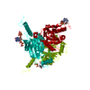
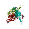
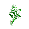
 Movie
Movie Controller


+ Open data

- Basic information
| Entry | Database: PDB / ID: 4ots | ||||||
|---|---|---|---|---|---|---|---|
| Title | Crystal Structure of isolated Operophtera brumata CPV18 | ||||||
 Components Components | Polyhedrin | ||||||
 Keywords Keywords | VIRAL PROTEIN / microcrystals / polyhedra / occlusion body | ||||||
| Function / homology | Cypovirus polyhedrin, Cypovirus 1 type / Cypovirus polyhedrin protein / GTP binding / ATP binding / ADENOSINE-5'-TRIPHOSPHATE / GUANOSINE-5'-TRIPHOSPHATE / Polyhedrin Function and homology information Function and homology information | ||||||
| Biological species |  Operophtera brumata cypovirus 18 Operophtera brumata cypovirus 18 | ||||||
| Method | X-RAY DIFFRACTION / SYNCHROTRON / MOLECULAR REPLACEMENT / Resolution: 1.704 Å | ||||||
 Authors Authors | Stuart, D.I. / Sutton, G.C. / Axford, D. / Ji, X. | ||||||
 Citation Citation | Journal: Acta Crystallogr.,Sect.D / Year: 2014 Title: In cellulo structure determination of a novel cypovirus polyhedrin. Authors: Axford, D. / Ji, X. / Stuart, D.I. / Sutton, G. | ||||||
| History |
|
- Structure visualization
| Structure viewer | Molecule: MolmilJmol/JSmol |
|---|
- Downloads & links
-Download
| PDBx/mmCIF format | 4ots.cif.gz | 147 KB | Display | PDBx/mmCIF format |
|---|---|---|---|---|
| PDB format | pdb4ots.ent.gz | 125.3 KB | Display | PDB format |
| PDBx/mmJSON format | 4ots.json.gz | Tree view | PDBx/mmJSON format | |
| Others |  Other downloads Other downloads |
-Validation report
| Arichive directory | https://data.pdbj.org/pub/pdb/validation_reports/ot/4otsftp://data.pdbj.org/pub/pdb/validation_reports/ot/4ots | HTTPS FTP |
|---|
-Related structure data


-Links
 PDBj
PDBj- Assembly
| Deposited unit | 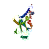
| |||||||||||||||
|---|---|---|---|---|---|---|---|---|---|---|---|---|---|---|---|---|
| 1 | 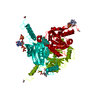
| |||||||||||||||
| Unit cell |
| |||||||||||||||
| Components on special symmetry positions |
|
-Components
-Protein , 1 types, 1 molecules A
| #1: Protein | Mass: 28301.373 Da / Num. of mol.: 1 Source method: isolated from a genetically manipulated source Source: (gene. exp.) Operophtera brumata cypovirus 18 / Plasmid: pOPIN / Production host:   Spodoptera frugiperda (fall armyworm) / Strain (production host): SF9 / References: UniProt: Q30C70 Spodoptera frugiperda (fall armyworm) / Strain (production host): SF9 / References: UniProt: Q30C70 |
|---|
-Non-polymers , 5 types, 245 molecules 


| #2: Chemical | ChemComp-CL / Chloride Mass: 35.453 Da / Num. of mol.: 1 / Source method: obtained synthetically / Formula: Cl Mass: 35.453 Da / Num. of mol.: 1 / Source method: obtained synthetically / Formula: Cl |
|---|---|
| #3: Chemical | ChemComp-GTP / Guanosine triphosphate Mass: 523.180 Da / Num. of mol.: 1 / Source method: obtained synthetically / Formula: C10H16N5O14P3 / Comment: GTP, energy-carrying molecule*YM Mass: 523.180 Da / Num. of mol.: 1 / Source method: obtained synthetically / Formula: C10H16N5O14P3 / Comment: GTP, energy-carrying molecule*YM |
| #4: Chemical | ChemComp-ATP / Adenosine triphosphate Mass: 507.181 Da / Num. of mol.: 1 / Source method: obtained synthetically / Formula: C10H16N5O13P3 / Comment: ATP, energy-carrying molecule*YM Mass: 507.181 Da / Num. of mol.: 1 / Source method: obtained synthetically / Formula: C10H16N5O13P3 / Comment: ATP, energy-carrying molecule*YM |
| #5: Chemical | ChemComp-MG /  Mass: 24.305 Da / Num. of mol.: 1 / Source method: obtained synthetically / Formula: Mg Mass: 24.305 Da / Num. of mol.: 1 / Source method: obtained synthetically / Formula: Mg |
| #6: Water | ChemComp-HOH / WaterMass: 18.015 Da / Num. of mol.: 241 / Source method: isolated from a natural source / Formula: H2O |
-Experimental details
-Experiment
| Experiment | Method: X-RAY DIFFRACTION / Number of used crystals: 20 |
|---|
- Sample preparation
| Crystal | Density Matthews: 1.6 Å3/Da / Density % sol: 23.08 % |
|---|---|
| Crystal grow | Temperature: 301 K / pH: 7.5 Details: Crystals formed naturally within the cytoplasm and were purified from cells, pH 7.5, temperature 301K |
-Data collection
| Diffraction | Mean temperature: 100 K |
|---|---|
| Diffraction source | Source: SYNCHROTRON / Site: Diamond  / Beamline: I24 / Wavelength: 0.9686 Å / Beamline: I24 / Wavelength: 0.9686 Å |
| Detector | Type: DECTRIS PILATUS 6M / Detector: PIXEL / Date: Jul 28, 2011 / Details: mirrors |
| Radiation | Monochromator: double crystal Si(111) / Protocol: SINGLE WAVELENGTH / Monochromatic (M) / Laue (L): M / Scattering type: x-ray |
| Radiation wavelength | Wavelength: 0.9686 Å / Relative weight: 1 |
| Reflection | Resolution: 1.7→72.686 Å / Num. all: 19343 / Num. obs: 19343 / % possible obs: 97.2 % / Observed criterion σ(F): 1.34 / Observed criterion σ(I): 1.34 |
| Reflection shell | Highest resolution: 1.7 Å |
- Processing
| Software |
| ||||||||||||||||||||||||||||||||||||||||||||||||||||||||
|---|---|---|---|---|---|---|---|---|---|---|---|---|---|---|---|---|---|---|---|---|---|---|---|---|---|---|---|---|---|---|---|---|---|---|---|---|---|---|---|---|---|---|---|---|---|---|---|---|---|---|---|---|---|---|---|---|---|
| Refinement | Method to determine structure: MOLECULAR REPLACEMENT / Resolution: 1.704→27.473 Å / SU ML: 0.12 / σ(F): 1.34 / Phase error: 11.18 / Stereochemistry target values: ML
| ||||||||||||||||||||||||||||||||||||||||||||||||||||||||
| Solvent computation | Shrinkage radii: 0.9 Å / VDW probe radii: 1.11 Å / Solvent model: FLAT BULK SOLVENT MODEL | ||||||||||||||||||||||||||||||||||||||||||||||||||||||||
| Refinement step | Cycle: LAST / Resolution: 1.704→27.473 Å
| ||||||||||||||||||||||||||||||||||||||||||||||||||||||||
| Refine LS restraints |
| ||||||||||||||||||||||||||||||||||||||||||||||||||||||||
| LS refinement shell |
| ||||||||||||||||||||||||||||||||||||||||||||||||||||||||
| Refinement TLS params. | Method: refined / Origin x: -20.9564 Å / Origin y: 8.6787 Å / Origin z: -34.0137 Å
| ||||||||||||||||||||||||||||||||||||||||||||||||||||||||
| Refinement TLS group | Selection details: all |
